The Muscles of the Digits
The remaining muscles of llie two limbs are, primarily, muscles of the digits, and are attached either to the basi-digital (metacarpal or metatarsal) bones, or to the phalanges, though they may acquire secondary connections with bones of the tarsus or carpus. The plan upon which they are arranged, when they are most completely developed, will be best understood by commencing with the study of their insertion in any one of those digits which possesses a complete set; such, for example, as the fifth digit of the manus, or little finger, in Man and the higher Primates.On the dorsal aspect this digit presents: first, attached to the base of its metacarpal bone, the tendon of a distinct muscle, the extensor carpi ulnaris. Secondly, spreading out over the phalanges into an aponeurosis, which is principally attached to the first and second, is a tendon belonging to another muscle, the extensor minimi digit. Thirdly, entering the same expansion is one tendon of the extensor communis digitorum.
On the ventral aspect there are: first, attached to the base of the metacarpal, the tendon of a distinct muscle, the flexor carpiulnaris ; secondly, arising from the sides and ventral face of the metacarpal, and inserted into either side of the base of the proximal phalanx, two muscles, the interossei; thirdly, inserted into the sides of the middle phalanx by two slips, a tendon of the flexor perforatus; and fourthly, passing between these two slips, and inserted into the base of the distal phalanx, a tendon of the flexors perforans. Thus there are special depressors, or flexois, for each segment of the digit. There appear, at first, to be but three elevators, or extensors, but, practically, each segment has its elevator. For the tendons of the extensor Communis and extensor minimum digit! are attached to the middle and the proximal phalanges; and the distal phalanx is specially elevated by the tendons of two little muscles, which, in Man, are usually mere subdivisions of the interossei, and pass upward, joining the extensor sheath, to be finally inserted into the distal phalanx.
The fifth digit of the pes, or little toe, sometimes presents the same disposition of muscles, namely:
On the dorsal aspect: first, the peronceus tertius for the metatarsal bone; secondly, one tendon from the extensor digitorum brevis, but this last is commonly absent in Man; thirdly, one tendon from the extensor digitorum longus.
On the ventral aspect: first, the Peronoeus brevis, attached to the base of the metatarsal ; secondly, two interossei; thirdly, a perforated flexor; and fourthly, a perforating flexor, like those of the manus. The divisions of the interossei, wliich send tendons to the extensor sheath on the dorsum of the digits of the foot in Man, are hardly distinct from the ventral divisions of those muscles.
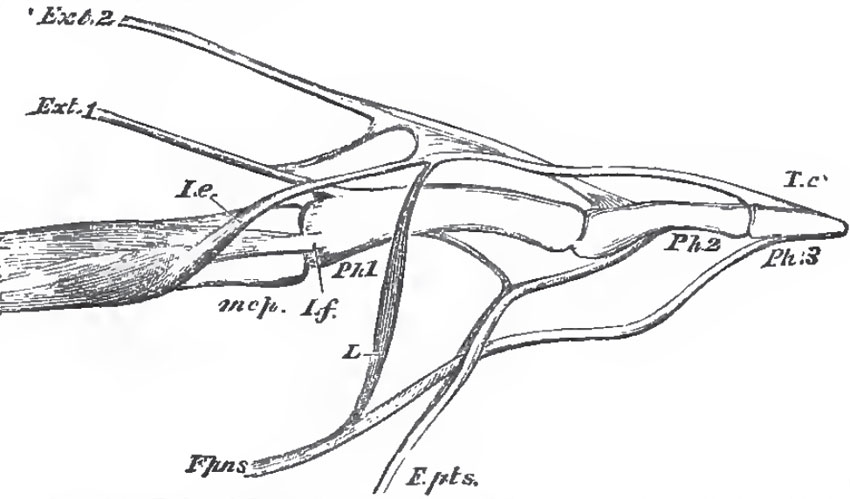 |
| Fig. 17. - Part of the middle digit of the manus of an Orang with the flexors and extensors of the phalanges: mcp, metacarpal bone; Ph. 1, Ph. 2, Ph. 3, the three phalanges; Ext 1, the deep long extensor tendon from the extensor indicis; Ext. 2, the supeificial long extensor tendon from the extensor communis; I. e, the interosseous short extensor; I.f, the interosseous short flexor; F.pns., the deep long flexor (perforans); F. pts., the superficial long flexor (perforatus). |
Finally, a lumbricalis muscle proceeds from the tendon of the perforating flexor, on the pre-axial side of the digit, to the extensor sheath.
None of the other digits of the manus, or of the pes, has a greater number of muscles than this; in fact, all the others have fewer muscles, some of those enumerated being suppressed. What are often regarded as muscles special to man, such as the extensor proprhis indicis and extensor minimidigiti, are only reuiains of muscles which are more fully developed in lower mammals, and send tendons to all four of the ulnar digits.
Only the pollex has an opponens (I have seen an opponens in the hallux of an Orang.) Only the pollex and hallux have adductors and adductors. Some of the digits luck one or more of the ventral, or of the dorsal, muscles.
The correspondence between the muscles which have been mentioned, at their insertion in the digits, is clear enough, but some difficulties present themselves when the muscles are traced to their origins.
In Man, the flexors and extensors of the digits (except the interossei) of the fore-limb arise in part from the humerus, and in part from the bones of the forearm, but not within the manus. On the contrary, none of the flexors and extensors of the digits of the pes arise from the femur, while some of them arise within the pes itself. The origins of the muscles seem to be, as it were, higher up in the fore-limb than in the hind-limb. Nevertheless, several of the muscles correspond very closely. Thus, on the dorsal aspect, the extensor ossis metacarpi pollicis passes from the post-axial side of the proximal region of the antebrachium obliquely to the trapezium and the metacarpal of the pollex, just as its homologue, the tibialis anticus, passes from the post-axial side of the upper part of the leg to the entocuneiform and the base of the metatarsal of the hallux; the two muscles correspond exactly. But the extensors of the phalanges of the pollex, and the deep extensors of the other digits of the manus, arise on the same side of the antebrachium, below the extensor ossis metacarpi pollicis; while, in the leg, one of the deep extensors of the hallux, and all those of the other digits, arise still lower down, viz., from the calcaneum.
Not less remarkable is the contrast between the more superficial sets of extensors in the two limbs. In the fore limb, proceeding from the pre-axial to the post-axial side, the following extensor muscles arise from the external or preaxial condyle of the humerus: the extensor carpi radialis Iongus to the base of the second metacarpal; the extensor carpi radialis brevis to the base of the third metacarpal; the extensor communis digitorium to the four ulnar digits; the extensor minimi digiti to the fifth digit; the extensor carpi ulnaris to the base of the fifth metacarpal. In the hind-limb, there are no homologues of the first two of these muscles. The homologue of the extensor communis is the long extensor, which arises, not from the femur, but from the fibula. The peroneus tertius, (This muscle, which lies altogether on the dorsal face of the hind-limb, and which 1 have seen only in Man, should not be confounded, as it often is, with one or more muscles, the peronnei 3tii, 4ti, et, 5ti digiti, which are very often developed in other Mammali, but arise on the ventral face of the fibula, and send their tendons below the external malleolus to the extensor sheaths of the fifth fourth and even third digits.) passing from the dorsal face of the fibula to the fifth metatarsal, is the only representative of the extensor carpi ulnaris.
On the ventral aspect of the human fore-limb, two deep flexors arise from the radius, ulna, and interosseous membrane, and run parallel with one another, though disconnected, to the digits. These are, on the pre-axial side - the Flexor pollicus longus, to the distal phalanx of the pollex; and the Flexor digitorum perforans, to the distal phalanges of the other digits.
In the hind-limb, two homologous muscles, the flexor halucis longus and the flexor digitorum perforans, arise from the tibia and fibula and interosseous membrane, and their tendons are distributed to the distal phalanges of the digits. But, before they divide, the tendons become connected together in such a way that many of the digits receive tendinous fibres from both sources.
In the fore-limb, there are no other deep flexors, but the internal, or post-axial, condyle of the humerus gives origin to a number of muscles. These, proceeding from the pre-axial to the post-axial side, are the flexor carpi radialis to the base of the second metacarpal; the palmaris longus to the fascia of the palm; the flexor perforatus digitorum to the middle phalanges of the four ulnar digits; the flexor carpi ulnaris to the base of the fifth metacarpal. The sesamoid, pisiform bone is developed in the tendon of the last muscle.
The only muscle which exactly corresponds with any of these, in the hind-limb, is the plantaris; which, in Man, is a slender and insignificant muscle proceeding from the outer (post-axial) condyle of the femur to the plantar fascia - and answers to the palmaris longus. In many quadrupeds, as the Rabbit and Pig, the plantaris is a large muscle, the tendon of which passes over the end of the calcaneal process ensheathed in the tendo achillis, and divides into slips, which become the perforated tendons of more or fewer of the digits.
The flexor carpi radialis is also roughly represented by the tibialis posticus - a muscle which passes from the tibia and interosseous membrane to the entocuneiform, and therefoie differs in insertion, as well as in origin, from its analogue m the fore-limb. The flexor perforatus digitorum of the foot takes its origin sometimes from the calcaneum; sometimes, in part from the calcaneum, and in part from the perforating flexor; or it maybe closely connected with the tendons of the plantaris. The peroneus brevis represents the flexor carpi ulnaris by its insertion, but it arises no higher than the fibula, and has no sesamoid.
Two most important muscles yet remain to be considered in the leg. The one of these is that which is inserted by the tendo achillis into the calcaneum, and arises by four heads, two from the condyles of the femur (called gastrocnemius), and two from the tibia and fibula (called soleus). The other muscle is the peroneus longus, arising from the fibula, passing behind the external malleolus, and then crossing the foot to the base of the metatarsal of the hallux.
The latter muscle does not appear to have any representative in the fore-limb. The gastrocnemius and soleus may possibly represent the crural part of the perforated flexor, since, in many of the Vertebrata, the tendo achillis is but loosely connected with the calcaneum, and passes over it into the plantar fascia and the perforated tendons. A peculiar adductor muscle of the hallux in Man and Apes is the transversalispedis, which is inserted into the basal phalanx of the hallux, and arises from the distal ends of the metatarsals of the other digits. The muscle sometimes has an analogue in the manus.
Support our developers


