The Non-Dediduate Mammalia
A large number of the non-deciduate Mammalia are oonveniently comprehended under the title of the Ungulata, though it may be open to question whether the group thus named represents a single order, or more than one.In all the Ungulata the placenta is either diffuse, that is to say, the villi are scattered evenly over the surface of the chorion; or it is cotyledonary, in which latter case, the villi are accumulated in distinct patches on the chorion. These patches are called cotyledons.
All Ungulata have milk-teeth, succeeded vertically by teeth of the permanent set. The teeth consist of enamel, dentine, and cement, and the grinders have broad crowns, with tuberculated, ridged, or folded enamel.
Clavicles are never present. The limbs have not more than four complete digits. The ungual phalanges are clothed in obtuse horny sheaths, which are commonly very thick and go by the name of hoofs. Upon these the weight of these quadrupeds is usually supported, whence they have been called ungaligrade. Some few, however, rest the weight of the body upon the under surfaces of the phalanges, or are digitigrade. The metacarpal and metatarsal bones are elongated, and take a vertical, or much inclined position.
In the female, the mammae are either few in number, when they are inguinal in position; or numerous, when they are disposed in two rows along the abdomen.
The intestiie is very generally provided with a caecum of considerable size.
The cerebral hemispheres always exhibit convolutions, which are usually very numerous; and, when the brain is viewed from above, the surface of the cerebellum is largely uncovered.
The Ungulata are divisible into the Perissodactyla and the Artiodactyla, though it is probable that the attempt to define these groups will break down with the increase of our knowledge of fossil forms.
 |
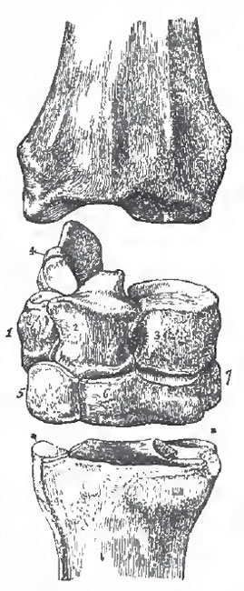
| Fig. 93. - A, Front aspect of the left tarsus of a Horse.-1. Calcaneum. 2. Astragalus. 3.
Naviculare. 4. Ectocuneiform. 5. Caboides. B, Posterior aspect of the left metatarsus of a Horse.-1. The metatarsal of the third digit 2, 3. The metatarsals of the rudimentary digits. |
In the skull, the tympanic bone is small; and, as in sundry other Mammals, the root of the pterygoid process of the sphenoid is perforated by an aperture or canal.
The posterior premolar teeth are, generally, very like the molars. The stomach is simple, and the caecum exceedingly large.
The teats are inguinal, or situated in the groin. When the head is provided with horny appendages, they are entirely epidermal and devoid of a bony core; and they are placed in the middle line of the skull.
The Perissodactyla consist of the existing families Equidae, Rhinocerotidae, and Tapiridae, and of the extinct Palaeotheridae and Macrauchenidae.
a. The Equidae, or Horses and Asses, have one toe on each foot-the third-much longer and larger than the rest. The latter are represented only by their metacarpal or metatarsal bones, the inner and outer toes being absent, or represented by mere ossicles (as rudiments of their metacarpals or metatarsals) in all existing Equidae. But, in the extinct Hipparion, the second and fourth digits were complete, though small and like dew-claws; while the miocene Anchitherium, which most nearly approaches the Palaeotheridae, has the lateral toes much larger, and taking their share in supporting the weight of the body.
 |
| Fig. 94. - A, right fore-foot of a Horse.-1. Radius. 2. Groove In the front face of the radius us. 3. Scaphoides. 4. Lunare. 5. Cuneiforme. 6. Pisiforme. 7. Magnum. 8. Unciforme. 9. Meticarpale, iii. 10. Metacarpale, iv. 11. Sesamoid bones in the ligaments
at the back of the motacarpo-phalangeal articulation. 12. Proximal phalanx (fetter-bone). 13. Widdle phalanx (coronary). 15. Distal phalanx (coffin-bone). 14. Sesamoid bono in the tendon of the flexor perforans (called "navicular" by Veterinarians). |
The molar teeth present an outer wall, which is bicrescentic in transverse section; and two inner ridges, which are curved more or less inward and backward, and correspond respectively with the anterior and the posterior crescents of the outer wall. The valleys may be more or less completely filled up with cement, which also coats the tooth. The incisors are similar in form in each jaw, and in Equus and Hipparion their crowns present a wide and deep median cavity, formed by a fold of the enamel.
These are the distinctive characters of the Equidae. It may be useful to add some special details respecting the anatomy of the Horse as a familiar example of the perissodactyle group.
The Horse has seven cervical vertebrae, twenty-four dorsolumbar (eighteen or nineteen of which are dorsal), five sacral, and about seventeen caudal vertebrae. The atlas has very wide lateral processes, the faces of which look obliquely downward and forward, and upward and backward. The centra of the other cervical vertebrae are much elongated, strongly convex in front, and correspondingly concave behind. The neural spines are obsolete in all but the seventh. The liga-mentum nuchae is a great sheet of elastic tissue, which extends from the spines of the anterior dorsal vertebrae to the occiput, and is fixed, below, into the neural arches of the cervical vertebrae.
In the dorsal region, the opisthocoelous character of the centra of the vertebrae gradually diminishes, though the anterior face of the centrum of the last lumbar is still distinctly convex. The spines of these vertebrae increase in length to the fourth or fifth. The spine of the sixteenth is vertical, those in front inclining backward, and those behind a little forward.
In none of these vertebrae do the prezygapophyses bend round the postzygapophyses of the vertebra in front, as is often the case in the Artiodactyla. The transverse processes of the penultimate, and of the last, lumbar vertebras present concave facets upon their posterior margins, which articulate with convex facets developed upon the anterior margins of the last lumbar and first sacral vertebrae respectively.
 |
| Fig. 95. - A cervical vertebra of a Horse.-1. The rudimentary spine. 2, 3. Tie pre- and post-zygapophyses. 5. The convex anterior face of the centrum. 9. Its concave posterior face. 6. 7. The transverse processes and rudimentary ribs. |
The tympanic bulla is not very large, and is rugose inferiorly. It is not anchylosed with the surrounding bones. The post-tympanic process of the squamosal does not approach the post-glenoidal process of the same bone, below the meatus auditorius.
The proper mastoid process is distinct, but short. There is a long and strong paramastoid developed from the ex-occipital.
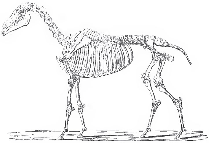 |
| Fig. 96. The Skeleton of the Horse. |
The structure of the limbs of the Horse is such as might be expected from its preeminent cursorial powers.
That excessive development of the epidermis which gives rise to a nail takes place, in the Horse, not only upon the dorsal surface of the terminal joint of the digit, but upon its ventral surface and sides, and thus produces a hoof.
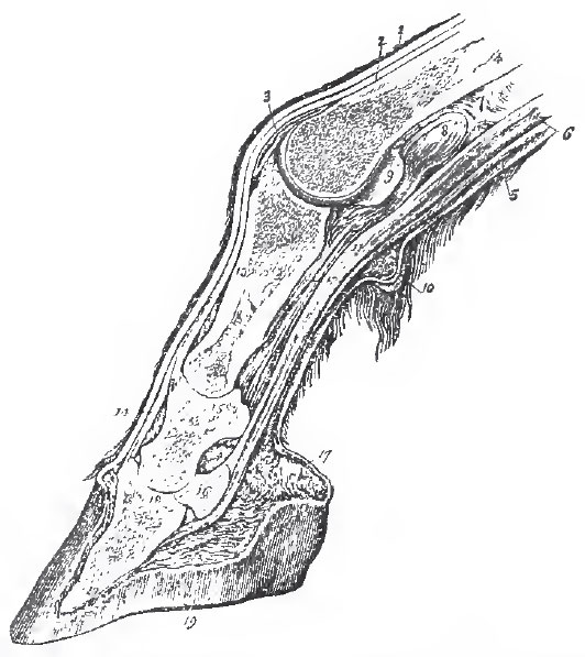 |
| Fig. 97. - Longitudinal median section of the foot of a IIerse.-13, 14, 18. The three phaslanges. 16. The navicular sesamoid. 5. The flexor perforatus. G. Tho flexor perforans 19. The hoof. |
The scapula is long and narrow; the low spine has no acromion; the coracoid process is small, and there is no clavicle.
 |
| Fig. 98. - Front view of the red carpas of a Horse.-1. Cunelforme 2. Lunare. 3. Scaphoides. 4. Pisiforme. 5. Unciforme. 6. Magnum. 7. Trapezoides. |
The pollax and the fifth digit are supressed, or represented only by minute nodules of bone, and the only complete digit is the third; the second and the forth being represented only by the splint-like metacarpal bones. The third metacarpal, which is somewhat flattened from before backward, is nearly symmetrical in itself. Careful observation, however, shows the inner moiety to be rather the broader.
There are two large sesamoid bones (the greater sesamoids) developed in the ligaments which connect the metacarpal with the basal phalanx; and one transversely-elongated sesamoid gives attachment to the tendon of the perforating flexor, and lies upon the ventral aspect of the joint between the middle and the distal phalanx.
The ossa innominata are elongated, and their long axes, on the length of which depends the proportional size of the "quarter" of a Horse, form an acute angle with the spine. The crests of the ilia are wide and directed transversely, and the symphysis pubis is very long.
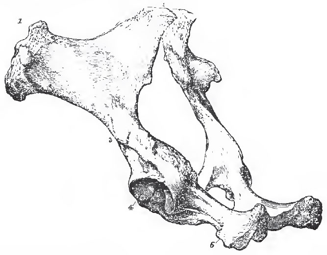 |
| Fig. 99. - The ossa innominata of a Horse viewed from the left side and behind.-1. The crest of the ilium. 2. The surface by which it articulates with the sacrum. 4. The acetabulum. 6. The ischium. |
The proximal end of the fibula is reduced to a mere rudiment; its shaft is not represented by bone; and its distal end is anchylosed with the tibia, and has the appearance of being an external malleolar process of that bone. The distal end of the tibia presents two deep, obliquely-directed concavities, which correspond with the convexities of the astragalus.
There are six or seven tarsal bones, according as the entoand meso-cuneiform bones remain distinct or become anchylosed. The astragalus (Fig. 93 A, 94 B) is extremely characteristic. It presents two convex ridges separated by a deep fossa, and directed obliquely from behind and within, forward and outward, to the tibia; and it has a nearly flat distal face, not borne upon any distinct neck, which articulates almost wholly with the naviculare, presenting only a very small facet to the cuboid.
The naviculare and the ecto-cuneiform are peculiarly broad and flattened in form (Fig. 93 A, 94 B).
The metatarsus and digits repeat the arrangements of the fore-limb; but the principal metatarsal is more slender in its proportions, and is flattened from side to side rather than from before backward (Fig. 93 B, 94 B).
As might be expected, the principal peculiarities of the muscular system of the Horse are to be observed in the limbs.
The serratus magnus and the levator anguli scapulae (which really form one muscle), together with a sterno-scapurlaris, form the great sling already mentioned, by which the weight of the forepart of the body is transmitted to the anterior extremities. The power of abduction is hardly needed by a purely cursorial animal; hence the deltoid is reduced to its scapular portion, which is very small. On the other hand, the pro-and re-tractors, the flexors and extensors, are well developed. The supra-and infra-spinatus are large. There is a great cephalo-humeralis, answering to the clavicular portions of the human sternomastoid and of the deltoid, which run into one another, in consequence of the total absence of the clavicle. The anterior portion of the sternomastoid is fixed to the mandible, and thus becomes "sternomaxillary."
The latissimus dorsi and teres muscles are very large, as are the flexors and extensors of the antibrachium.
The supinators and pronators are wanting; but there is a distict extensor minimi digiti, the tendon of which unites with that of the extensor communis. Radial and ulnar extensors of the carpus are also present. The, flexor perforatus has only a single tendon, which splits, and is attached, as usual, to the sides of the middle phalanx. The flexor perforans also has only a single tendon, which pierces the former, and is inserted into the lesser sesamoid and the distal phalanx.
The intorossei of the third digit are represented only by the ligaments which connect the greater sesamoid bones with the metacarpal, and in which a few muscular fibres are sometimes found. There are said to be two others, one for each lateral metacarpal, and a lumbricalis.
In the hind-limb, the femoral muscles are in the Horse the same as in Man, but enormously developed. There is no tibialis anticus, peronaeus longus, or brevis, nor any tibialis posticus.
The extensor longus digitorum has a head which arises from the external condyle of the femur; there is a simple extensor brevis.
The flexor hallucis and flexor digitorum perforans unite into the single perforating flexor tendon for the distal phalanx; while the perforated tendon is the termination of that of the plantaris, which passes over a pulley furnished by the calcaneum.
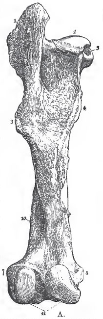 |
| Fig. 100. - The left femur of a Horse, posterior view - 1. Head 2. Great grand trochanter 3. Third trochanter 4. Lesser trochanter 5. Pit for round ligament. 10. Fossa. 11. Condyles. |
In the mandible, the structure of the molars and the resulting pattern are quite different. The outer wall presents two convex surfaces separated by a longitudinal depression, and thus reverses the conditions observable in the upper molars. The result of the wear of this is, necessarily, two crescents, the concavities of which are turned inward. A vertical pillar, longitudinally grooved on its inner face, is developed on the inner face of the tooth at the junction of the anterior and posterior crescents, and gives rise to a deeply bifurcated surface when worn. A second smaller pillar appears in connection with the inner face of the posterior end of the outer wall.
Thus the grinding surface of the upper molars may be represented by four crescents with two inner pillars; and that of the lower molars by two crescents with two inner pillars. The upper crescents are concave outward; the lower concave inward; and by this arrangement, together with the unequal wear of the dentine, enamel, and cement, a permanently uneven triturating surface is secured.
As is the general rule among Mammals, the first permanent molar is the first permanent tooth which appears (unless the eruption of the inner incisor be contemporary with it), and it comes into place and use long before the deciduous molars are shed and replaced by the premolars. Hence, when the last premolar comes into place as a fresh and unworn tooth, the first molar, which lies next to it, is already considerably worn. This disparity of wear is maintained for a long time, and furnishes a very useful means of distinguishing the last premolar from the first molar in the adult, when, as in the Horse, the premolars and molars are very similar.
The first deciduous molar usually falls out when the first premolar appears, and is not replaced; but it is occasionally retained. AH the other milk-teeth have successors, and there are three permanent molars. Consequently the dental formula of the adult Horse is i. 3.3/3.3 c. 1-1/1-1 p.m. 3.3/3.3 m. 3.3/3.3=40.
The permanent canines are the last teeth to be fully developed, and, in the mare, they do not often make their appearance. The upper canines are distant from the outer incisors, while the lower canines are quite close to them. In both jaws there is a wide interval, or diastema, between the canines and the premolars.
The deep valley of the incisor teeth becomes filled up with masticated matter, and thus the dark "mark" is produced. As the incisors wear down, the mark changes its form in consequence of the differences in the transverse section of the valley at different points; and eventually, when the wear has extended beyond the bottom of the valley, it disappears. The presence or absence of the "mark" thus serves as an indication of age. The structure and patterns of the grinding surfaces of the permanent molars are essentially the same as those of the milk-molars; but the enamel becomes more or less plaited; and, at an advanced period of life, the development of the long teeth is completed by the formation of roots. It is important to notice that the last molar of the Horse is not more complex in its structure than the other molars, and that the last milk-molar is not more complex than the premolar which succeeds it.
The alimentary canal of the Horse is about eight times as long as the body. The stomach, simple in its form, presents a cardiac and a pyloric division, which are sharply distinguished by the dense epithelium which lines the inner surface of the former.
The caecum is enormous, having fully twice the volume of the stomach. There is no gall-bladder. A cartilage is developed in the septum of the heart. There is no Eustachian valve, and only one anterior cava remains. The aorta divides immediately after its origin into an anterior and a posterior trunk; the latter becomes the thoracic aorta; the former is the source of the arteries for the head and the anterior ex tremities, giving off first the left subclavian, and then as an "innominata" supplying the right subclavian and the carotids.
The trachea divides into only two bronchi, no accessory bronchus being given off to the right lung. In the brain the following points are worthy of notice: The medulla oblongata presents corpora trapezoidea. The flocculi do not project at the sides of the cerebellum, and the vermis and lobes of the cerebellum are unsymmetrically convoluted. The cerebral hemispheres are elongated and subcylindrical, and do not overlap the cerebellum when the brain is viewed from above, The sulci are very deep, and separate numerous gyri, upon the upper and outer surfaces of the hemispheres. The uncinate gyrus (or natiform protuberance) and the region which answers to the insula are not hidden by the overlapping of the convolutions in the lateral aspect of the brain. The Sylvian fissure is indicated. The corpus callosum is large, and the anterior commissure is of moderate size. The posterior cornu of the lateral ventricle is wanting.
Large air-sacs are connected with the Eustachian tubes.
The testes pass into a scrotum, but the unguinal canal remains permanently open.
The prostate is single. Cowper's glands are present, and there is a large uterus masculinus. The large penis is sheltered within a prepuce and is retracted by a special muscle, which arises from the sacrum.
The uterus is divided into two cornua, and the vagina of the virgin mare is provided with a hymen. The period of gestation is eleven months. The yelk-sac of the foetus is small and oval. The allantois spreads over the whole interior of the chorion and covers the amnion, which is vascular. The minute villi which it supplies with vessels are evenly scattered over the whole surface of the chorion.
The existing Equidae, are naturally restricted to Europe, Asia, and Africa; and are distinguished into the Horses, which have homy patches on the inner sides of both pairs of limbs-above the wrist in the fore-limb and on the inner side of the metatarsus in the hind-limb; and the Asses, which possess such callosities only on the fore-limbs.
Fossil remains of Equidae and abundant in the later tertiary deposits of Europe, Asia, and the Americas; but the group is not known to be represented earlier than the miocene, or later eocene, epoch.
The Equidae are among the very few groups of Mammalia, the geological history of which is sufficiently well known, to prove that the existing forms have resulted from the gradual modification of very different ancestral types. The skeleton of the older pliocene and newer miocene Hipparion very closely resembles that of an Ass, or a moderate-sized Horse. There is a curious depression on the face in front of the orbit, somewhat like that which lodges the "larmier" of a stag (traces of which are observable in some of the older species of Equus); otherwise the cranium is altogether like that of a Horse. Again, the shaft of the ulna is very slender, but it is larger than in the Horse, and is distinctly traceable throughout its whole length although firmly anchylosed with the radius. The distal end of the fibula is so completely anchylosed with the tibia, that, as in the Horse, it is difficult to discern any trace of the primitive separation of the bones. But, as has been already mentioned, each limb possesses three complete toes-one strong, median, and provided with a large hoof, while the two lateral toes are so small that they do not extend beyond the fetlock-joint. In the fore-limb, rudiments of the first and fifth toes have been found.
The teeth are exceedingly like those of the Horse, but the crowns of the molars are shorter; and, in the upper jaw, that which, in the true Horses, is a large fold of the inner face of tho tooth becomes a detached pillar. The smaller plications of the enamel are also more numerous, close-set, and complicated. On the outer face of the lower milk-molars there is a column such as exists in the Stags. Of this a rudiment exists, as a fold, in the corresponding teeth of the existing Horse.
In the genus Anchitherium, all the known remains of which are of older miocene (and, perhaps, newer eocene) age, the skeleton in general is still extraordinarily like that of a Horse. The skull, however, is smaller in proportion than in the Horse, and the jaws are more slender. The hindermost molar tooth is situated farther back under the orbit, and the orbit itself is not completely encircled by bone, as it is in the Horses and Hipparions.
The shaft of the ulna is stouter than in Hipparion, and is less closely united with the radius. The fibula appears, at any rate in some cases, to have been a complete though slender bone, the distal end of which is still closely united with the tibia, though much more distinct than in the Hipparions and the Horses. In some specimens, however, the middle of the shaft seems to have been incompletely ossified. Not only are there three toes in each foot, as in Hipparion, but the inner and the outer toes are so large that they must have rested upon the ground. Thus, so far as the limbs are concerned, the Archithericum is just such a step beyond the Hipparion, as the Hipparion is beyond the Horse, in the direction of a less specialized quadruped. The teeth are still more divergent from the Equine type. The incisors are smaller in proportion, and their crowns lack tbe peculiar pit which characterizes those of Equus and Hipparion. The first grinder is proportionately much larger, especially in the upper jaw, and like the other six has a short crown and no thick coat of cement. The pattern of their crowns is wonderfully simplified. The fore and hind ridges run with but a slight obliquity across the crown, and the pillars are little more than enlargements of the ridges, while in the lower jaw these pillars have almost disappeared. But the foremost of the six principal grinders is still somewhat larger than the rest, and the posterior lobe of the last lower molar is small, as in the other Equidae.
In all those respects in which Anchitherium daparts from the modern Equine type, it approaches that of the extinct Palaetheria; and this is so much the case that Cuvier considered the remains of the Anchitherium with which he was acquainted to be those of a species of Palaeotherium.
b. In the Rhinocerotidae the second, third, and fourth toes are nearly equally developed in both the fore-and the hind feet.
The dental formula is i. 1.1/1.1 or i. 0.0/0.0 p.m. 4.4/4.4 m. 3.3/3.3 But the teeth differ from those of the Horse in many other respects besides the number of the incisors and the absence of canines. Thus, the upper incisors differ greatly in form from those which are situated in the lower jaw; and, in some species, incisors are absent. Their crowns are not folded as in the Horse. The peculiarities of the grinding teeth will be mentioned below.
The skin is very thick and may be converted into a jointed armor; the hair is scanty. The upper lip is much produced and is very flexible. In some species one, or sometimes two, horns are attached in the middle line to the nasal or frontal bones. But these horns are formed, as it were, by agglomeration of a great number of hair-like shafts.
The distal phalanges of the tridactyle feet of the Rhinoceros are invested by small hoofs; but these do not entirely support the weight of the body, which rests, in great measure, upon a large callous pad developed from the under face of the metacarpal and metatarsal regions; these are much shorter than in the Horse.
The dorso-lumbar vertebrae are twenty-two or twenty-three, of which twenty are dorsal. There are four sacral and twenty-two caudal. The cervical vertebrae, as in the Horse, are strongly opisthocoelous, and the transverse processes of the last lumbar articulate with those of the penultimate lumbar and with the sacrum.
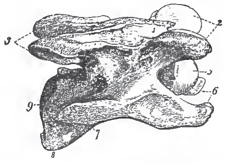
The skull differs from that of the Horse in the absence of any frontal or zygomatic processes in consequence of which the orbit and temporal fossa form one cavity. The nasals are immense, and are separated from the premaxillae by a wide extent of the maxilla on each side. The premaxillae are relatively small and reduced to little more than their palatine portions. The glenoidal surface of the mandible is transverse and convex. The squamosal sends down an immense postglenoidal process, which is longer than either the post-tympanic or the paramastoid. It unites with the post-tympanic to form a kind of false auditory meatus, in the absence of any proper ossified canal of that kind. The periotic and the tympanic bones are anchylosed, the tympanic being a mere irregular hoop of bone. The pars mastoidea is completely hidden by the junction of the short post-tympanic with the long par amastoid. The hinder margin of the bony palate is opposite the middle of the antepenultimate molar.
The mandibular condyle is transverse and convex. The perpendicular portion of the ramus is large, and the coronoid process ascends slightly above the condyle. In a vertical and longitudinal section of the skull, the form of the cerebral cavity is seen to be similar to that of the Horse. The inner and outer tables of the bony roof of the skull are separated by great air-cavities.
The spine of the scapula has no acromion, but gives off a strong recurved process from the middle of its length.
The radius and ulna are complete, but are anchylosed.
The carpus has the eight ordinary bones. In the manus the digits ii., iii., iv., are complete, and a bony tubercle articulated with the outer facet of the cuneiforme represents digit v. The digit iii. is largest and longest, and its phalanges are symmetrical in themselves; those of the digits ii. and iv. are not symmetrical in themselves. The terminal phalanges have somewhat the form of the coffin-bone of the Horse.
The ilia have wide, transversely-directed crests, as in the Horse. The femur is provided with a very strong third trochanter. The tibia and the fibula are complete, and the tarsus has the ordinary seven bones. The pulley of the astragalus is not very deeply grooved, and is hardly at all oblique. The facet for the cuboid is very small. The metatarsals resemble the metacarpals in their number and symmetry, but there is no rudiment of the fifth.
In some species of Rhinoceros there are 3.3/2.2 incisors in tlie milk detention, and 1.1/2.2 or 1.1/1.1 incisors in the permanent dentition. In the latter the upper incisors are large, long-crowned teeth, very unlike the lower ones, of which it seems probable that only one pair, in any case, are permanent teeth. In some Rhinoceroses, as has been already stated, the adult is devoid of incisor teeth.
There are no canines in either dentition. Of the four milkmolars, the first, as in the Horse, is smaller than the others, and is not replaced. The structure of both the upper and the lower molars is substantially the same as in the Horse, but the roots are developed much sooner; the laminae of the upper molars take a much more transverse direction; the laminae of the upper molars do not develop pillars, though accessory crests may be developed from the two faces of the posterior lamina; the lower molars have no pillars; and the cement does not fill up the valleys between the wall and the laminae.
The cardiac division of the simple, though large stomach, is lined by a white callous epithelium, as in the Horse. The small intestine presents large processes or tags, half an inch long or more, upon which the true villi are borne. The caecum is very large, and the colon enormous. There is no gall-bladder. The heart and brain are very similar to those of the Horse.
The male can hardly be said to have a scrotum, as the testes lie close to the abdominal ring. A prostate, vesiculae seminales, and Cowper's glands, are present. The long penis has a mushroom-shaped glans, and the animal is retromingent. The cornua uteri are proportionately longer than in the mare. The teats are two and inguinal in position. The characters of the foetal membranes and the nature of the placentation are unknown.
At the present day the genus Rhinoceros is confined to Africa and Asia. The African species all have two horns, a nearly smooth skin, and the adult has no incisors. The Asiatic species have one horn only (except that of Sumatra, which has two). The skin is marked out by deep folds into shields, and the adults have well-developed incisors.
Rhinoceroses are known in the fossil state as far back as the miocene epoch. R. tichorhinus with the nasal septum ossified, and a covering of long woolly hair, inhabited Europe and Asia during the cold of the glacial epoch., R. incisivus had four digits in the manus, and larger incisor teeth than any existing species. R. hexaprotodon had more numerous incisors than any other species.
c. In the Tapiridae there are four toes on the front-foot, though the ulnar digit does not reach the ground. The hind foot has three toes.
The dental formula is i. 3.3/3.3 c. 1.1/1.1 p.m. 4.4/3.3 m. 3.3/3.3.
The molar teeth each present two transverse, or slightly oblique ridges, connected by a low wall externally.
The skin is soft and hairy, and the muzzle and snout are prolonged into a short proboscis.
The Tapirs have twenty-three or twenty-four dorso-lumbar vertebrae, of which nineteen or twenty are usually dorsal. The centra of these vertebrae, and the transverse processes of the last lumbars, have the same peculiarities as those of the Horse and Rhinoceros. There are seven sacral and about twelve caudal vertebra. The skull is partly Rhinocerotio, partly Equine, in its characters. Thus there is a sagittal crest- the post-tympanic processes are large, but they are not so long as the paramastoids, and they do not unite with the postglenoidal processes beneath the meatus. In these respects the Tapir is Horse-like, but in the following it is more Rhinocerotio.
Thus the tympanic is quite rudimentary; the post-glenoidal process is larger than in the Horse; the orbit is not separated from the temporal fossa; the nasals are widely separated from the premaxillae; the premaxillae are very small, and are early anchylosed.
The hinder margin of the osseous palate is opposite the anterior edge of the penultimate molar. The mandibular rami unite in a very long symphysis; the ascending portion of the ramus is large, and projects-backward with a convex edge in a remarkable manner. There is a high coronoid process.
In the fore-limb, the scapula has no acromion, and the coracoid is a mere tubercle. The supraspinous fossa is very much larger than in the Horse or Rhinoceros. The radius and the ulna are complete, but not movable upon one another. Although, by the completion of the fifth digit, in addition to the second, third, and fourth, there are four digits in the manus, the Perissodactyle character is manifested by the fact that the third is longest, and symmetrical in itself, while the others are asymmetrical. The femur has a strong third trochanter; the fibula is complete; the astragalus more Rhinocerotio than Equine. There is no trace of a hallux, but the fifth digit of the pes appears to be represented by an osseous rudiment.
In the presence of the full complement of incisors and canines the Tapir is more Horse-like than Rhinocerotic, but is still very peculiar; for the outer upper incisors are larger than the canines, while the outer lower incisors are much smaller than the canines, and are apt to fall out at a certain age. The canines, are still more closely approximated to the incisors than in the Horse, especially in the lower jaw, and, consequently, the diastema is very large. The six posterior molars in the upper jaw, and the five posterior molars in the lower, present nearly the same structure. There is a low outer wall with two slightly-marked concavities (in the maxillary teeth) or convexities (in the mandibular teeth) on its outer face. From this two ridge-like laminae run inward and a little backward across the crown of the tooth. The valleys are broad and shallow, and the coat of cement very thin. The molar tooth of the Tapir thus represents the plan of structure common to the Perissodactyle in its simplest form. Deepen the valleys, increase the curvature of the wall and lamina, give the latter a more directly backward slope; cause them to develop accessory ridges and pillars, and increase the quantity of cement; and the upper molar of the Tapir will gradually pass through the structure of that of the Rhinoceros to that of the Horse.
In the anterior upper premolar (or milk-molar?) the anterior moiety of the crown is incompletely developed. In the anterior lower premolar the anterior basal process, which exists in all the molars, is excessively developed, so that the crown of the tooth assumes the bicrescentic pattern of the Rhinoceros's lower grinder. This probably indicates the manner in which the Tapiroid form of inferior molar is converted into the Rhinocerotic, or Equine, form.
The stomach is simple and oval, the cardiac and pyloricorifices being closely approximated. The caecum is proportionally smaller than in the Horse or Rhinoceros. There is no gall-bladder. The heart is devoid of a septal bone and of a Eustachian valve. There is only a single vena cava anterior, and the aorta divides into an anterior and a posterior trunk. There is no third bronchus. No distinct scrotum is present There are vesiculae seminales and prostatic glands, but no Cowper's glands. The placentation is diffuse. The teats are two, and inguinal.
There are two or three species of Tapir at present living in South America and one in Southwest China, Malacca, and Sumatra. The genus Tapirus has been found fossil in Europe in rocks of miocene age. The closely-allied extinct genera Lophiodon (and Coryphodon?) carry the Tapiridae back through the eocene epoch.
d. The Palaeotheridae.-These are all extinct animals, the remains of which are found in the older tertiary rocks; and which are closely allied, on the one hand, with the Horses and, on the other, with the Tapirs.
The type of the family, Palaeotheridium, resembles the Tapir in most respects, but has only three digits in the manus as well as in the pes. The dental formula, however, is i. 3.3/3.3 c. 1-1/1-1 p.m. 4.4/4.4 m. 3.3/3.3. The diastema is smaller than in the Tapir, and the patterns of the grinding teeth of both jaws are more like those of the Rhinoceros.
e. The Macrauchenidae. - The genus Macrauchenidae. is also extinct form, which occurs in later tertiary or quaternary deposits in South America.
The feet are tridactyle, and the dental formula is i. 3.3/3.3 c. 1.1/1.1 p.m. 5.5/4.4 m. 3.3/3.3. The teeth are disposed in a nearly continuous series. The crowns of the incisors present a deep fossa, as in the Equidae. The molars are in part Equine, in part Rhinocerotic in character. The skull is, on the whole. Equine, but the nasal bones are very short and Tapiroid. The vertebrae of the long nock are extraordinarily similar to those of the Camelidae, and especially of the Llamas.
Support our developers


