Supercoils in closed DNA
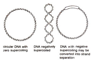
Fig. 25.19. Supercoiling of DNA leading lo a twisted duplex, which may be undone by strand separation in a region.
Fig. 25.19. Supercoiling of DNA leading lo a twisted duplex, which may be undone by strand separation in a region.
The supercoils may be negative (as found in vivo), when they are in a direction opposite to the clockwise turns of right handed DNA. This will actually relieve the torsion and will become underwound or even single stranded in a region. This helps in unwinding of DNA for replication, etc. The positive supercoils lead to overwinding (superhelical), which can be created in vitro, but does not occur in nature. Supercoiling is often controlled by some enzymes and is described by parameters like linking number, twisting number and writhing number (for details, consult Lewin's 'Genes V').
Support our developers


