The Basics of Mitosis
In mitosis, the daughter nuclei each retain the same number of chromosomes as the parent nuclei; in meiosis, by reduction division, the daughter nuclei each have half the chromosome number of the parent. We thus speak of the diploid number and the haploid number of chromosomes. Common practice is to represent the haploid number with the letter N. The diploid number, therefore, is 2N. In a general sense, all non reproductive cells in a body have the same number of chromosomes-that is, the diploid number. This is clear because a body grows in size by mitosis and cell division. All members of the same species, with few exceptions, have the same number of chromosomes. Many know that the human chromosome number is 46 (2N = 46), the haploid number being 23. The chromosome number of cabbages is 18 (2N = 18); corn has 20 chromosomes; sunflowers have 34; and plums have 48. In the animal kingdom, cats have 38 chromosomes, dogs have 78, and crayfish have approximately 200; In the plant kingdom, the fern Ophiuglussum vulgatum has 500 chromosomes.The process of mitosis, once initiated, is a continuous flow. It is convenient, however, to designate the various stages with names. Figure 3-1 shows the events in animal mitosis, provided for purposes of comparison. The student who is already familiar with these details may wish to proceed to the section on plant mitosis.
The top left illustration in figure 3-1 shows a cell in which there is no evidence of nuclear division. This is the interphase of mitosis. While this is sometimes called the resting stage, resting it is not. In fact, much synthetic activity is occurring. The Golgi bodies, endoplasmic reticulum, and ribosomes all increase to provide enough for two cells, should cell division occur. The DNA is replicated, and protein is synthesized. Mitochondria and chloroplasts also reproduce themselves at this time. Considering the entire time lapse from the beginning of interphase to the beginning of the next interphase, approximately 90 percent of the time of mitosis is spent in interphase.
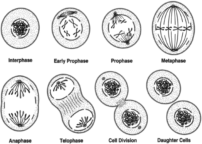 |
Figure 3-1 The events of mitosis in animal cells: interphase, showing an intact nuclear
membrane and two centrioles lying side by side: early prophase, when the centrioles
begin to move apart and a spindle begins to form in prophase, the chromosomes
replicate but remain attached at the centromeres; metaphase, only the centromeres of the
chromosomes lie on the equatorial plate; anaphase; telophase, when the chromosomes
have reached the centrioles at the poles; cell division, the process of mitosis is having been
completed; and daughter cells. |
The next stage in mitosis is prophase. During prophase, the centrioles begin to move apart, the nuclear membrane disappears, and the nuclear material forms what first appear to be fine threads and then clearly takes the form of chromosomes. The chromosomes are then free in the cytoplasm. In prophase, the chromosomes replicate, and their number doubles. The example in figure 3-1 shows a cell with a diploid number of 4 (2N = 4). When chromosomes replicate, giving the appearance of splitting down the middle, they do so faithfully, particle by particle. The products of this process are two members of the .same composition. These members remain temporarily attached to each other at a point called the centromere, or kinetochore.
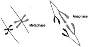 |
Figure 3-2 Chromosomes replicate during prophase. At left, they are shown at metaphase. They then part beginning at the point of the centromeres and go into anaphase. |
The chromosomes migrate to a position along an equatorial plate, and the centrioles continue their movement, until they lie at opposite poles of the cell, one on each side of the centrally placed chromosomes. This is called metaphase. Careful examination reveals spindle fibers running from one centriole to the other and from the centriole to the centromere of the replicated chromosomes.
The chromosomes which had been attached next become disjoined and begin to move away from the equatorial plate and toward the centrioles. At this time, it appears that the spindle fibers attached to the centromeres contract, thus pulling the chromosomes toward the centrioles. This is called anaphase (see figure 3-2).
The migration of chromosomes continues until they reach the centrioles, at which time the cell is in telophase. During this stage, the chromosomes seem to lose their organization, becoming indiscernible as chromosomes: the centrioles divide: and new nuclear membranes are formed. The nucleolus also reforms at this time, apparently at a specific locus of a chromosome called the nucleolar organizer. Two examples are shown in figure 3-3.
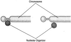 |
Figure 3-3 The nucleolar organizer can nestle against a chromosome at two possible positions. |
This marks the completion of mitosis, and the daughter nuclei return to interphase. If cell division is to occur, it will become evident at this time.
Support our developers


