 |
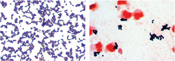
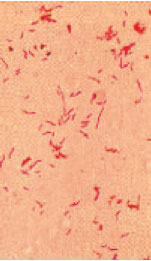
| Plate 1 Staphylococcus aureus in a Gram-stained smear from a colony growing on agar medium (left) and from the sputum of a patient with staphylococcal pneumonia (right). The organisms are gram-positive spheres, primarily in grapelike clusters. The pink cells in the right-hand photo are neutrophils. |
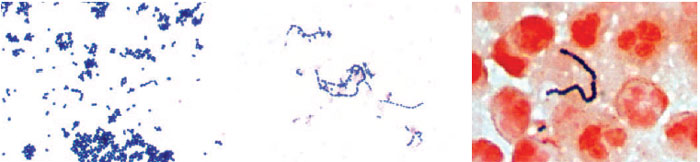 |
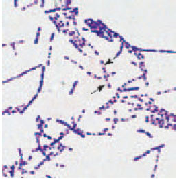
| Plate 2 Streptococcus pyogenes in Gram-stained smears. From a culture plate (left), the gram-positive organisms appear singly, in chains, and in clumps. In broth culture media (center), the characteristic long chains are seen. In a smear from an abscess (right), the organisms are primarily gram-positive cocci in long chains. |
 |
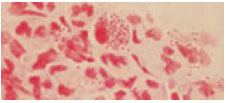
| Plate 3 Streptococcus pneumoniae in Gram-stained smears. The organisms from a colony growing on agar medium (left) are gram positive and lancet shaped and appear in pairs and short chains. In a Gram stain of cerebrospinal fluid from a patient with pneumococcal meningitis (right), the organisms are mostly diplococci. The capsule (arrow) can be seen around some bacteria, outlined by the pink proteinaceous material of the fluid. |
 |
| Plate 4 Neisseria gonorrhoeae in a Gram-stained smear from a male urethral exudate appear as gramnegative, bean-shaped diplococci. |
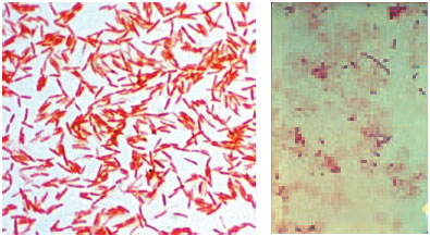 |
| Plate 5 Gram-negative bacilli (Klebsiella pneumoniae) in a Gramstained smear from an agar colony (left) and a patient’s blood culture (right). In the blood specimen, the organisms are pleomorphic, varying in length from coccobacillary to filamentous. |
 |
| Plate 6 Curved, spiral, gram-negative bacilli (Campylobacter jejuni) in a Gram stain from culture. Some bacteria line up to form spirilla like chains. |
 |
| Plate 7 Spirochetes (Treponema pallidum) appear black (arrows) in a skin preparation stained with a silver stain. |
 |
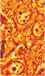
| Plate 8 Bacillus spp. are gram-positive bacilli with endospores. Endospores appear as clear areas within the vegetative bacterial cell (arrows). |
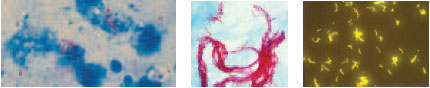 |
| Plate 9 Mycobacterium tuberculosis in an acid-fast stain of sputum (left). The acid-fast bacilli appear as red, beaded rods against a blue background (×1,000). Strains of M. tuberculosis often form ropy “cords” (center), which is considered an indication of virulence. When stained with a fluorescent dye, acid-fast bacilli fluoresce brightly against a dark background (right, × 400). |
 |
| Plate 10 The quellung reaction. The halo around the cells is the pneumococcal capsule, which appears to swell when the cells are treated with pneumococcal antiserum. |
|


