 |
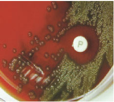
| Plate 31 Optochin test. A zone of inhibition forming around a disk containing optochin (P disk) identifies this organism presumptively as Streptococcus pneumoniae. |
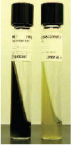 |
| Plate 32 Esculin reaction. The Enterococcus sp. on the left has hydrolyzed esculin with resulting blackening of the medium. The Streptococcus sp. on the right does not hydrolyze esculin. |
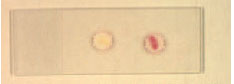 |
| Plate 33 PYR test. The appearance of a red color at the completion of the test indicates that the organism on the right is PYR positive. |
 |
| Plate 34 Satellite test. Colonies of Haemophilus influenzae, which requires both X and V factors, grow only around a Staphylococcus aureus streak on a blood agar plate. The blood provides the needed X factor (hemin) and the staphylococcus, V factor (a coenzyme, NAD). |
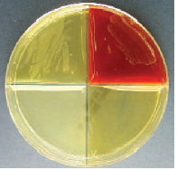 |
| Plate 35 Haemophilus ID Quad Plate inoculated with Haemophilus influenzae. The organism grows only on the top two quadrants, which contain media supplemented with X and V factors (left) and 5% blood and V factor (right). |
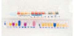 |
| Plate 36 Rapid bacterial identification. In the Enterotube II (top) and API strip (bottom), many reactions are tested simultaneously allowing definitive organism identification within 24 hours. |
 |
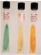
| Plate 37 Phenol red agar slants containing glucose, maltose, sucrose, and fructose inoculated with oxidasepositive, gram-negative diplococci. Only the first tube (glucose) shows a positive reaction, indicating the organism is Neisseria gonorrhoeae. |
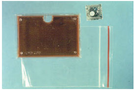 |
| Plate 38 The JEMBEC plate is used primarily when genital specimens for culture must be transported long distances to the microbiology laboratory. After the white CO2-generating tablet (top right) is placed in the well in the rectangular culture plate, the plate is sealed in the plastic zip-lock bag. CO2 accumulates, providing the appropriate atmosphere for growth of Neisseria gonorrhoeae. |
 |
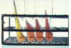
| Plate 39 Growth of Mycobacterium spp. on Lowenstein- Jensen slants. The green tube on the left is uninoculated. The tube in the center has the characteristic dry, heaped, and rough growth of M. tuberculosis. The tube on the right shows the yellow pigmented growth of the photochromogen, M. kansasii. |
|


