The cell
Content
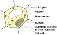
Without the use of a microscope, the horticulturist will not be able to see cells, since they are very small (about a twentieth of a millimetre in size). They are very complex and scientific studies continue to discover more of the organization displayed in this fundamental unit. A simple, unspecialized cell of parenchyma (see Figure 6.2) consists of a cellulose cell wall and contents (protoplasm) enclosed in a cell membrane, which is selective for the passage of
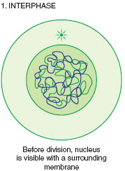
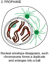
Suspended in the jelly-like cytoplasm are small structures (organelles) each enclosed within a membrane and having specialized functions within the cell. In all tissues, the cell walls of adjoining cells are held together by calcium pectate (a glue-like substance which is an important setting ingredient in 'jam-making'). Some types of cell (e.g. xylem vessels) do not remain biochemically active, but die in order to achieve their usefulness. Here, the first wall of cellulose becomes thickened by additional cellulose layers and lignin, which is a strong, impervious substance. The cell is made up of two parts, the nucleus and the cytoplasm. The nucleus coordinates the chemistry of the cell. The long chromosome strands that fill the nucleus are made up of the complex chemical deoxyri-bonucleic acid, usually known as DNA. In addition to its ability to produce more of itself for the process of cell division, DNA is also constantly manufacturing smaller but similar RNA (ribonucleic acid) units, which are able to pass through the nucleus membrane and attach themselves to other organelles. In this way, the nucleus is able to transmit instructions for the assembling, or destruction, of important chemicals within the cell. There are six main types of organelle in the cytoplasm. The first, the vacuole is a sac containing dilute sugar, nutrients and waste products. It may occupy the major volume of a cell, and its main functions are storage and maintaining cell shape. The ribosomes make proteins from amino acids. Enzymes, which speed up chemical processes, are made of protein. The Golgi apparatus is involved in modifying and storing chemicals being made in the cell before they are transported where they are required. Mitochondria release energy in a controlled way, by the process of respiration, to be used by the other organelles. The energy is transferred via a chemical called ATP (adenosine triphosphate). The meristem areas of the stem, root and flower have cells with the highest number of mitochondria in order to help the rapid cell division and growth in these areas. Plastids such as the chloroplasts are involved in the production of sugar by the process of photosynthesis, and in the short-term storage of condensed sugar (in the form of starch). Lastly, the endoplasmic reticulum is a complex mesh of membranes that enables transport of chemicals within the cell, and links with the plasmodesmata at the cell surface. Ribosomes are commonly located on the endoplasmic reticulum. The whole of the living matter of a cell, nucleus and cytoplasm, are collectively called protoplasm. Cell division
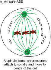
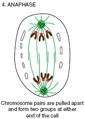
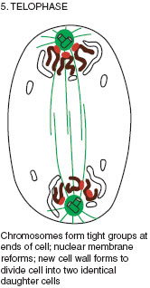
The genetic information in the nucleus is reproduced exactly in the new cells to maintain the plant’s characteristics. Each chromosome in the parent cell produces a duplicate of itself, thus producing sufficient material for the two new daughter cells (see Figure 6.3). A delicate, spindle-shaped structure ensures the separation of chromosomes, one complete set into each of the new cells. A dividing cell wall forms across the old cell to complete the division. |
|||||||||||||||||||||||||||||||||||||||||||||||||||||||
Support our developers


