Neurology of Sauropsida
In all the Sauropsida the cerebro-spinal axis is angulated at the junction of the spinal cord with the medulla oblongata, the latter being bent down toward the ventral side of the body. The region in which the nerves of the anterior and posterior extremities originate may be enlarged in reptiles, as in birds; but, in the former, the posterior columns of the cord remain parallel in the lumbar enlargement while, in the latter, they diverge and give rise to the sinus rhomboidals, which is a sort of repetition of the fourth ventricle, the dilated central canal of the spinal cord being covered merely by a thin membrane consisting chiefly of the ependyma and arachnoid.The brain (Fig. 90) fills the cavity of the skull in the higher Sauropsida, and presents a well-developed cerebellum; a mesencephalon divided above into two optic lobes; and relatively large prosencephalic hemispheres, which attain a considerable size in Crocodilia, and Aves, but never conceal the optic lobes. In Crocodilia the cerebellum presents a distinct vermis, with transverse fissures. In birds the latter are more distinct, and the lateral appendages of the cerebellum, or flocculi, become well defined, and are lodged, as in many of the lower Mammalia, in cavities of the side walls of the skull, arched over by the anterior vertical semicircular canal.
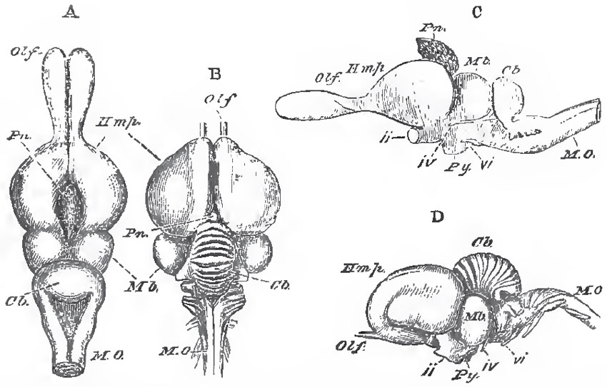 |
| Fig. 90. - A, C, the brain of a Lizard (Psammosaurus Bangalensis), and B, D, of a bird (Meleagris gallopuro, the Turkey), drawn as if they were of equal lengths. A, B, viewed from above; 0, D, from the left side. Olf., Olfactory lobes; Pn., Pineal gland; Hmp., cerebral hemispheres; Mb, optic lobes of the mid-brain; Cb., cerebellum; M.O., medulla oblougata; ii. iv., vi., second, fourth, and sixth pairs of cerebral nerves; Py.,pituitary body. |
Each prosencephalic lobe contains a lateral ventricle (continuous through the foramen of Munro with the third ventricle), which is little more than a fissure between the very thin inner wall of the lobe and its thick outer part, which contains the corpus striatum. The corpora striata are united by an anterior commissure, which is not of large size. The thinning of the inner wall of the lobes, from the margin of the foramen of Munro backward, which gives rise to the fissure of Bichat in the Mammalia, extends for a very short distance in the Sauropsida, even in birds.
The olfactory lobes are usually elongated, and contain ventricles continuous with those of the prosencephalic hemispheres.
In all Sauropsida the motor nerves of the tongue pass through a foramen in the exoccipital bone. Hence, twelve pairs of cranial nerves are present, except in the Ophidia, which possess no spinal accessory nerve.
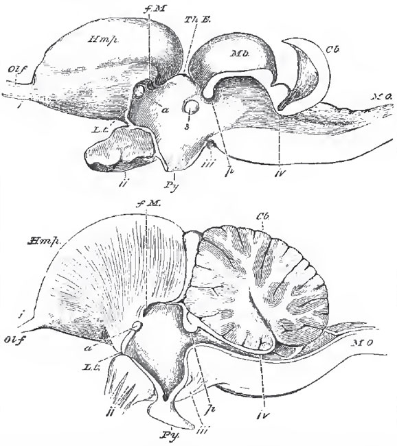 |
| Fig. 91. - The brains of a Lizard (Psammosaurus Bengalensis) and of a bird (Meleagrins gallopavo), in longitudinal and vertical section. The upper figure represents the lizard's brain; the lower (taken, like Fig. 90, B, D, from Carus's "Erlauterunga-Tafeln") that of the bird. The letters ss in the preceding figure, except L. t, lamina terminalis, or anterior wall of the tliird ventricle; f. M., foramen of Munro; a., anterior commissure; Th. E., thalamenephalon: s., soft commissure; p., posterior commissure; iv., indicates the exact point of exit of the fourth pair from that part of the brain which answers to the value of Vieussens. |
The sympathetic is well developed, except in the Ophidia, where it is not distinct from the spinal nerves, in the greater part of the trunk.
The Ophidia, many Sauria, and Aves, possess nasal glands, which, in birds, attain a large size, and lie more usually upon the frontal bone, or in the orbits, than in the nasal cavity.
Support our developers


