Myxomycetes
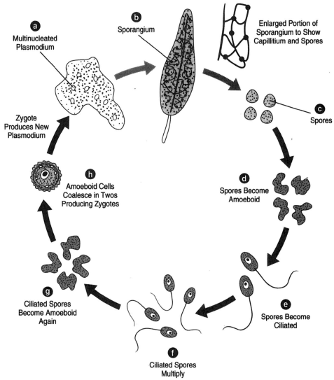
The organisms of Myxomycetes present a challenge in classification because during one part of the life cycle they exhibit animal-like characteristics, and during another part, they exhibit plantlike traits. A large, naked, noncellular mass of protoplasm possibly several inches in diameter and called a plasmodium can be found growing on damp forest debris. It may be variously colored yellowish, brown, green, and orange. It is capable of amoeboid movement and can glide slowly to get away from light. While it will shun light at one stage in its life cycle, it will move into the light at another stage. When a plasmodium reaches a certain maturity, sporangia grow upward from it, and it thus takes on a plantlike role.Stemonitis is an example. The vegetative phase of this slime mold can be found on rotting logs in the woods. It can be recognized by the creamy drops of liquid that it exudes to its surface. A large mass of protoplasm is found just under the drops. It is coenocytic and capable of amoeboid movement. In this way, it can avoid light by “flowing” to the underside of the log. This is the plasmodium of Stemonitis. The nuclei are diploid. Upon maturity, a number of fruiting bodies develop. Nuclei from the plasmodium migrate up into the fruiting bodies, which will become sporangia. Here, the nuclei undergo meiosis; the resulting spores are thus haploid. A wall called the peridium forms on the surface of the sporangium. Within the body of the sporangium, where the spores reside, is a mass of filamentous threads called the capillitium (see figure 17-1). The peridium disintegrates upon drying and releases the spores, which transform into amoeboid swarm spores. The swarm
 |
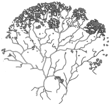
| Figure 17-2 An enlargement of the plasmodium showing protoplasmic streaming. |
spores soon undergo another change, developing cilia. In this form, they are able to go through a number of divisions. Then, after a short time, they lose their cilia and become swarm spores again. The swarm spores come together in twos, producing diploid zygotes, which, in turn, coalesce to produce a new coenocytic plasmodium. The coalescence of the swarm spores is due to the influence of the hormone acrasin. (Interestingly, an acrasinlike substance that accomplishes the same thing can be extracted from the urine of pregnant women.)
Microscopic examination of the plasmodium reveals a rhythmic flow of the cytoplasm; the cytoplasm flows for a moment, comes to a brief rest, and flows again in the opposite direction. While the cytoplasm flows forwards and backwards, the entire plasmodium can move continuously in one direction at a rate of nearly one inch per hour. It feeds on bits of organic material and bacteria along the way.
 |
| Figure 17-1 The life cycle of Stemonitis. (a) The multinucleated plasmodium and (b) The sporangium. At the right of the sporangium, a portion of sporangium much enlarged to show capillitium and some spores. (c) Spores, which become amoeboid, (d), and then ciliated, (e). The spores then multiply, (f), and again become amoeboid, (g). The amoeboid cells coalesce in twos, producing zygotes, (h), which, in turn, produce a new plasmodium. |


