T-2 Bacteriophage
 |
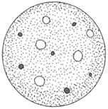
| Figure 7-1 A lawn of bacteria will show visible bare spots called plaques after scattered viruses that have touched those places have lysed and spread to neighboring cells. |
The T-2 phage particle, a virus, infects the common colon bacterium Escherichia coli. In experiments involving T-2 and E. coli, a Petri dish was inoculated with E. coli and incubated until the bacteria covered the entire surface of the dish. Next, a much diluted suspension of the T-2 virus was . spread on this bacterial “lawn.” After some time had passed, cleared spots called plaques appeared where the bacteria had been lysed (broken down); wherever a T-2 particle came into contact with the E. coli, the bacterium was “attacked by the T-2. This caused the bacterium to lyse. The T-2 particles, which replicated many times over in this way, spread to adjacent bacteria, which were then also lysed. The eventual result was a plaque large enough to be seen without the aid of magnification. Viruses of this kind, then, are intracellular parasites that, when grown inside a bacterium, result in the bacterium’s destruction.
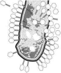
When the electron microscope came into use, it became possible to see things that could not be seen under a light microscope. It was even possible to see T-2 particles adsorbed (adhered) to the surface of an E. coli cell.
In 1952 Hershey and Chase learned that phage particles are susceptible to osmotic shock. They placed a suspension of phage in a concentrated salt solution. After several minutes, they suddenly diluted the solution. This caused the parts of the phage to separate. Ultracentrifugation then allowed the parts to be isolated and identified. Two components were found a protein and DNA.
 |
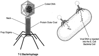
| Figure 7-2 At left, a T-2 phage particle showing the coiled DNA inside the protein outer coat; the necka;n d the prop organs. At right, an E. coli bacterial cell to which several T-2 particles have adsorbed. |
Phosphorus is a part of DNA but not a part of protein. Sulphur is a part of protein but not a part of DNA. In one series of experiments, Hershey and Chase made radioactive phosphorus, P32, a part of phage DNA to provide a traceable tag that would allow them to see where the phage DNA went when placed in contact with E. coli. Likewise, they used radioactive sulphur, s35 as a tag to show where the phage protein went. Some T-2 phages were first cultivated in a medium containing radioactive P32 and then were placed in E. coli. The T-2 phages of course grew in the E. coli. Although only one billionth of the phosphorus in the DNA was radioactive, this was enough. In this way, Hershey and Chase learned that most of the radioactivity was inside the E. coli cell; in other words, the DNA went inside the cell.
 |
| Figure 7-3 An electron micrograph showing a large number of T-2 virus particles attached to the outer surface of the bacterium E. coli. |
Next, Hershey and Chase cultivated T-2 phages in a medium containing radioactive s35, a constituent of protein. When the T-2 phages were placed in E. coli, the protein part of the phage remained outside of the bacterial cell.
Hershey and Chase’s findings coupled with electron microscope studies showed that the T-2 phage is composed of an outer protein portion and an inner DNA portion; and that the phage becomes adsorbed to the surface of E. coli and extrudes (forces) its DNA into the E. coli cell. The only part of T-2 that infiltrates the bacterial cell, then, is information. This again demonstrated that DNA is the genetic material. The electron microscope reveals the phage outer coat to take the form of a hexagonal head part, a tail portion, and some prop organs.
When the phage’s DNA enters the E. coli, it “takes over,” in a sense. By exploiting this energy source, many more phage particles are manufactured. When the phages increase to a certain number, the bacterial cell is lysed; the T-2 then spreads to adjacent E. coli cells. It is interesting to note that the components of the phage are not immediately assembled up on manufacture. The electron microscopes hows that at one point in the cycle, head parts, tail parts, and prop organs exist separately in the cell. Only at the last moment, just before lysis, are the complete phages assembled the DNA is taken into the head, the tail and prop organs are properly attached, and the bacterial cell is lysed. These events take approximately ten and one-half minutes from the time the T-2 first contacts the outside of the bacterium.


