The Cell Doctrine
The doctrine that all living things are made up of cells was published in 1839. This doctrine is credited to two men: Matthias Schleiden (1804-1881), a botanist, and Theodor Schwann (1810-1882), an anatomist. Though working independently, Schleiden and Schwann came to this conclusion at nearly the same moment.As it turns out the doctrine that all living things are composed of cells not tenable. Some things do not have a cellular organization. Yet, most organisms, both plant and animal, are constituted of cells; and cells, no matter what their sources, share certain characteristics. The cells of cabbages and giraffes, for example, have much in common. This suggests that the great diversity of life comes from a common beginning; and as focus is directed to the minute organelles that reside in cells, the differences fade even more to where such structures as mitochondria and Golgi bodies appear to be quite the same whatever their sources.
| Figure 2-3 Matthias Schleiden (810 4-1 881) contributed to the theory that all lifise composed of cells. (Illustration by Donna Mariano) |
As microscopes were improved, the structures contained within cells were revealed, and how these structures are involved in cell activity also became known. Our knowledge of cells has advanced along two fronts.On the one front, increasingly powerfuml icroscopes have allowedt he identification of the smaller aspects of the cell;on the other front, biochemical methods enabled the discovery of how these delicate microstructures are involved in metabolism.
 |
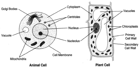
| Figure 2-2 At the left is a generalized animal cell showing mitochondrion, vacuole, nucleus, nucleolus, centrioles, and Golgi bodies. At the right is a plant cell, which conforms to the shape of a rigid cell wall. With the exceoption of the centrioles, which are generally not seen in plant cells, plant cell possesses the same organelles as are shown for the animal cell. Shown for the plant cell are chloroplasts, a large vacuole, and both primary and secondary cell walls, which lie outside of the cell membrane. |
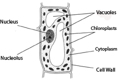
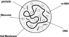
Figure 2-4 shows what can generally be seen with a light microscope. Now, consider for a moment the smallest imaginable cell having all the components necessary for the maintenance of life. A mycoplasm is an example. It is estimated that such cell needs more than of molecules in order to sustain and perpetuate its life. Such cells are smaller and less complex than the simplest bacteria. There is a delicate plasma membrane made of proteins and lipids. The cell is, of course, prokaryotic, that is, having no discrete nucleus. Recently, the electron microscope has allowed for magnification of such cells by much more than 100,000 diameters, giving us additional knowledge regarding cell structures. Following is a discussion of the mitochondria, Golgi body, endoplasmic reticulum, nuclear membrane, cell membrane, cell walls, chloroplasts, cilia, plastids, and vacuoles.
 |
Figure 2-4 Plant cell as seen through a light microscope: nucleolus, nuclceeulls wall,
cytoplasm, chloroplast, and vacuole. |
 |
Figure 2-5 A mycoplasm, the smallest possible cell containing all components
necessary for life: protein, unit membrane, m-RNA, DNA, and ribosome. |


