The Functions of DNA
 |
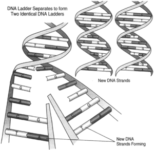
| Figure 6-7 Before DNA can replicate or act as a template for the manufacture of m-RNA, it must first uncoil. In forming a new DNA molecule, each spiral of the uncoiling helix has a newly made spiral wound around it. In this way, each new DNA molecule has a parent strand and a “newborn” strand. |
What does DNA do? The answer is two fold DNA can provide a pattern for the structuring of more DNA, which it must do when a cell divide; and DNA can provide a pattern for the making of RNA.
Self-Replication
To accomplish either of its functions-that is, self-replication or the patterning of m-RNA-the helices of DNA must uncoil. This is accomplished by the breaking of the hydrogen bonds that join the paired bases. In becoming uncoiled, each helicals trand forms a companion strand, and the two strands again become coiled (see figure 6-7). Specifically, a freed adenine base can attract a nucleotide containing thymine; likewise, a freed cytosine base can attract a nucleotide containing guanine. As the new strand of DNA is completed, it becomes entwined with its parent. There are then two new double helices, each of which is made up of an older strand and a newly formed strand. In this manner, DNA is increased for subsequent mitoses.
Patterning of RNA
When the structure of DNA was determined, it became clear that the bases along the helix represented coded information, that the sequencing of the bases represented a code, and that the coded information carried instructions to be used in creating proteins. DNA, however, does not leave the nucleus; it must therefore send instructions to the cytoplasm by way of a messenger. That messenger is RNA.
While most of it lies outside of the nucleus and in the cytoplasm, small amounts of RNA are present in chromosomes. RNA is manufactured in the nucleus. RNA differs from DNA in three ways. As mentioned earlier, the sugar in RNA is ribose rather than deoxyribose; and the base uracil is used in its structure in place of the thymine found in DNA. Finally RNA molecules are single rather than double-stranded (although an RNA molecule may appear double stranded when it bends back on itself).
There are three known kinds of RNA: ribosomal RNA, messenger RNA (m-RNA), and transfer RNA (t-RNA). DNA controls what goes on in a cell by sending a messenger; the messenger molecule is m-RNA. One of the functions of DNA is to provide a template for the formation of m-RNA.
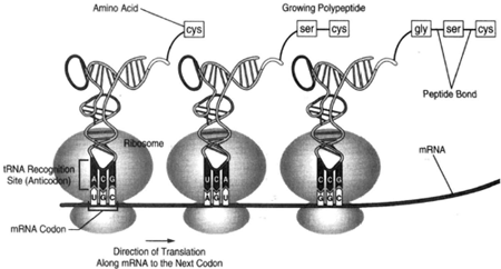
In performing this function-that is, the patterning of m-RNA-the helices of DNA again uncoil; but only one DNA strand plays a role in the fabrication of the messenger molecule. Recall that in m-RNA, uracil is used in the place of the thymine found in DNA. The new m-RNA molecule, which has been fabricated on the DNA template, moves out of the nucleus through the micropores of the nuclear membrane. It then becomes linked with the ribosomes that lie along the course of the endoplasmic reticulum. An electron microscope reveals the ribosomes to be like two beads: one smaller and the other larger. The m-RNA moves through the ribosome (or the ribosome moves along the RNA molecule), and, in doing so, the bases pass through in their previously determined sequence. As shown in figure 6-8, the first three to meet the ribosome are uracil, guanine, and cytosine (UGC). Messenger RNA is a coded molecule having code words of three letters, and the ribosome is the manufacturing center for protein. The UGC triplet (or codon) of the m-RNA calls on the ribosome to place the amino acid cysteine on line. When the next codon, for example AGU, passes through the ribosome, the amino acid serine comes on line and is linked to the earlier placed cysteine. The amino acids are united by peptide bonds.
 |
| Figure 6-8 When m-RNA molecules leave the nucleus, they goto the ribosomes, which functions as protein assembly plants. The m-RNA molecules then impart to the ribosomes information implanted on the m-RNA molecules by contact with DNA; specifically, the m-RNA moleculest ell the ribosomes which particular amino acid to place on line in the assembly of protein. Here, the codon UGC has caused the ribosome to place the amino acid cysteine on line; and the codon AGU, having passed through the center ribosome, has caused serine to be connected to the previously placed cysteine. |


