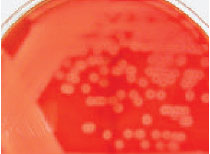
Plate 11Streptococcus pyogenes growing on a blood agar plate. The clear beta-hemolytic areas surrounding the punctate colonies are caused by a streptolysin enzyme.
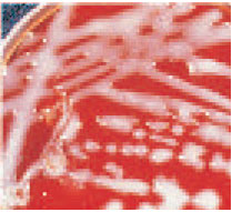
Plate 12 The large capsule of Klebsiella pneumoniae gives a mucoid appearance to colonies growing on agar plates.
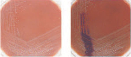
Plate 13Neisseria gonorrhoeae colonies on chocolate agar. The oxidasepositive
organisms become deep purple when a drop of oxidase reagent is
added to an area of the plate (right).
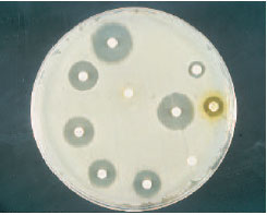
Plate 14 A disk diffusion antimicrobial susceptibility test. If the clear zones of growth inhibition around disks are of a certain diameter, the organism is susceptible to the antimicrobial agent in the disk.
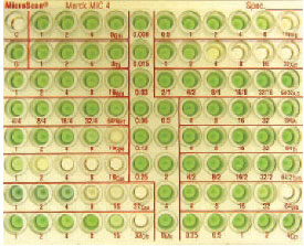
Plate 15 A microdilution susceptibility test of Pseudomonas aeruginosa. The green color signifies growth of the organism with production of its soluble green pigment. Growth occurs in the wells containing concentrations of antimicrobial agents to which the organism is resistant.
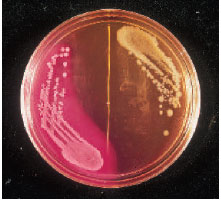
Plate 16 A MacConkey agar plate with Escherichia coli (pink, lactosefermenting colonies) growing on the left-hand side and a Salmonella sp. (colorless, lactose nonfermenting colonies) on the right.
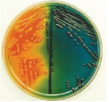
Plate 17Escherichia coli (left) and Salmonella sp. (right) on a Hektoen enteric agar plate. The lactosefermenting E. coli colonies appear yellow, whereas the Salmonella colonies appear black because of hydrogen sulfide production. Compare with reactions on colorplate 16 and note how selective and differential media display different organism characteristics.
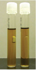
Plate 18 When Durham tubes are placed inside broth tubes, gas produced by fermentation of carbohydrates in the medium can be visualized as a bubble in the inner tube (left). The organism on the right does not produce gas when fermenting carbohydrates.
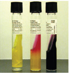
Plate 19 Kligler iron agar slants test for fermentation of glucose and lactose and the production of gas and hydrogen sulfide. The organism on the left ferments both glucose and lactose with gas production (bubbles in medium). The organism in the middle ferments glucose (yellow butt) but not lactose (pink slant). The organism on the right ferments glucose (with gas production) but not lactose, and blackens the agar as a result of hydrogen sulfide production. Reactions are similar on TSI slants.
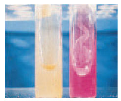
Plate 20 Urease test. The organism on the right produces the enzyme urease, which imparts the bright pink alkaline reaction to the urea agar slant.