 |
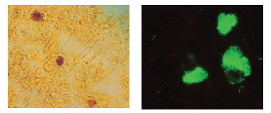
| Plate 40 Inclusions of Chlamydia trachomatis in cell culture. The glycogen-containing inclusions stain dark brown when the cells are treated with an iodine solution (left). |
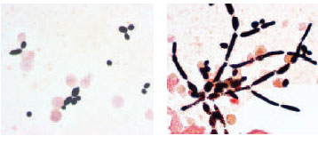 |
| Plate 41 Gram stain of Candida albicans cells isolated from the blood culture of a patient. At left the yeast cells are budding, and at right, they have formed long, filamentous, irregularly staining hyphae. |
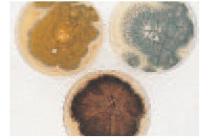 |
| Plate 42 Colonies of three Aspergillus species. Some molds may be recognized by the color of their spores (conidia). Clockwise from left: A. flavus (yellow), A. fumigatus (smoky gray-green), A. niger (black). |
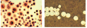 |
| Plate 43 Cryptococcus neoformans in cerebrospinal fluid from a patient with AIDS. In the Gram stain at left, yeast cells are seen to stain irregularly. The orange-staining halo around some cells is the Cryptococcus capsule. In the India ink preparation at right, the cryptococcal yeast cells are surrounded by a capsule that is demarcated by the suspension of charcoal particles in the India ink. |
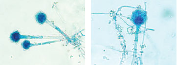 |
| Plate 44 Lactophenol cotton blue coverslip preparation from a slide culture of Aspergillus fumigatus. At low power (×100) magnification (left), three spore-bearing structures can be seen. At higher power (×400) magnification (right), the characteristics that permit genus and species identification are clearly visualized. |
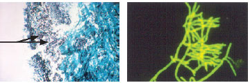 |
| Plate 45 Left: Methenamine silver stain of sinus biopsy (×100). All of the black material represents the invading fungus, Aspergillus flavus. Two of the characteristic sporebearing structures can be seen on the nasal cavity side (arrows). Right: A calcofluor stain of the biopsy material as seen under an ultraviolet light source (×400). |
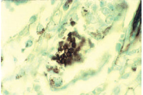 |
| Plate 46 Methenamine silver stain of cyst form of Pneumocystis carinii from lung biopsy of a patient with AIDS. |
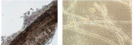 |
| Plate 47 KOH preparations of a hair and skin scales from patients with tinea capitis (ringworm of the hair) and tinea corporis (ringworm of the body). On the left, many round, reproductive spores of the dermatophyte fungus are seen surrounding the hair; the filamentous hyphae invade the hair shaft. On the right, the filamentous hyphae are seen invading skin scales throughout the preparation. |
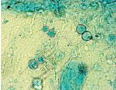 |
| Plate 48 Lactophenol cotton blue preparation of Blastomyces dermatitidis growing in culture of sputum. The characteristic thickwalled, broad-based budding yeast cells are seen at the top right and bottom left of the preparation. |
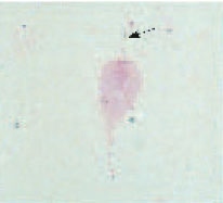 |
| Plate 49 Trichomonas vaginalis in a Gram stain of vaginal secretions. At least one flagellum (arrow) can be clearly seen. Most other parasites do not stain with the Gram stain. |
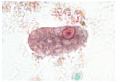 |
| Plate 50 A trophozoite of Entamoeba histolytica. The characteristic circular nucleus at the top right portion of the ameba has dark chromatin evenly distributed around its edges and a centrally placed karyosome. |
|


