Introduction to Ion Transport Across Biological Membranes
The movement of inorganic ions across biological
membranes of animals plays a central role in the perception
and integration of, and reaction to, environmental signals
by the organism. Examples include vision, the integration
and processing of this information (brain function),
and the reaction of the organism to this information, for instance,
muscle contraction (Fig. 1). Cells have the ability
to hydrolyze adenosine triphosphate (ATP). The energy
thus released is used to transport sodium and potassium
ions across the cell membrane against a concentration gradient.
This process establishes the transmembrane voltage
(V
m) of the cell membrane. The transmembrane voltage
is perturbed by the movement of inorganic ions (generally
Na
+, K
+, Cl
−, and Ca
2+) along the concentration gradient
and voltage difference across the cell membrane that occurs
in signal transmission between cells. There are many
different transmembrane channel-forming proteins, which
are activated by (1) a concentration gradient of inorganic
ions across the membrane, (2) the transmembrane voltage,
(3) the binding of specific ligands to a channel-forming
protein. (4) Some proteins use the energy liberated by
the hydrolysis of ATP to transport inorganic ions against a
concentration gradient. Only one example of each of these
various proteins will be mentioned. In each case, the protein
chosen is the one about which we have the most information.
The proteins that facilitate inorganic ion transport
across biological membranes are discussed in an order that
illustrates their function in the life of an organism.
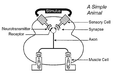 |
Figure 1 An environmental stimulus, for instance, light, activates
a protein-mediated reaction in the eye, leading to the transmembrane
flux of inorganic ions, a change in the transmembrane
voltage, and neurotransmitter release. The neurotransmitter diffuses
across a junction between the nerve terminal of the axon
and the cell body of the adjacent cell about 20–40 nm in length,
called synapse. The neurotransmitter binds to receptors in the
membrane of the postsynaptic cell. Excitatory neurotransmitters
( ) activate receptors that form cation-specific transmembrane
channels. Inhibitory neurotransmitters ( ) activate receptors that form cation-specific transmembrane
channels. Inhibitory neurotransmitters ( ) activate receptors that
form anion-specific transmembrane channels. Once the transmembrane
voltage of the cell is changed by a critical amplitude
and sign (by ~ +20 mV), an all-or-none process occurs. Transmembrane
Na+ and K+ channels in the axonal membrane open
transiently, resulting in an electrical signal that travels down the
axon and neurotransmitter is again secreted. This process repeats
itself and is terminated when the neurotransmitter is released
adjacent to receptors on the surface of muscle cells. In the
case of muscle cells, the receptor is the muscle nicotinic acetylcholine
receptor and the neurotransmitter acetylcholine. The voltage
change in the muscle cell membrane initiates muscle contraction.
(From Hess, G. P., and Grewer, C. (1998). “Methods in
Enzymology” (G. Marriott, ed.), Vol. 291, pp. 443–474, Academic
Press, New York.) The resulting flow of inorganic ions through the
membrane of the muscle cell results in a change of its transmembrane
voltage Vm and muscle contraction. ) activate receptors that
form anion-specific transmembrane channels. Once the transmembrane
voltage of the cell is changed by a critical amplitude
and sign (by ~ +20 mV), an all-or-none process occurs. Transmembrane
Na+ and K+ channels in the axonal membrane open
transiently, resulting in an electrical signal that travels down the
axon and neurotransmitter is again secreted. This process repeats
itself and is terminated when the neurotransmitter is released
adjacent to receptors on the surface of muscle cells. In the
case of muscle cells, the receptor is the muscle nicotinic acetylcholine
receptor and the neurotransmitter acetylcholine. The voltage
change in the muscle cell membrane initiates muscle contraction.
(From Hess, G. P., and Grewer, C. (1998). “Methods in
Enzymology” (G. Marriott, ed.), Vol. 291, pp. 443–474, Academic
Press, New York.) The resulting flow of inorganic ions through the
membrane of the muscle cell results in a change of its transmembrane
voltage Vm and muscle contraction. |


