Plant Mitosis
Thus far a clear distinction has been made between nuclear division (karyokinesis) and cellular division (cytokinesis)and although the intent at this point is to describe mitosis, it is fitting that cell division be drawn into the discussion.Plant mitosis is in many ways similar to animal mitosis. In animal cells, the cell membrane becomes constricted, being pinched in from the outside. This appears to be accomplished by micro filaments that behave in a manner similar to a purse string, pinching the cytoplasm into two parts. In plant cells, the first evidence of cell division is seen in the center of the cell.
Here, a cell plate forms and grows progressively outward. to meet the cell membrane. The cell plate arises from a line of vesicles produced by the Golgi bodies. An earlier statement regarding anaphase made reference to an
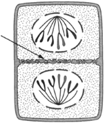 |
| Figure 3-4 When a plant cell divides, the new cell membrane that divides the two cells begins near the center and grows in an outward direction, as shown at |
impression that the spindle fibers contract to pull the chromosomes toward the centrioles at the poles. This, however, does not apply to a description of plant mitosis. For one thing, centrioles cannot be detected in most plant cells. In such plant cells, the chromosomes move toward the poles but not toward the centrioles. Even in those plant cells having centrioles, when the spindle fibers are cut and the centrioles removed by micro dissection methods, the events of mitosis go on in a normal manner. If spindle fiber contraction were responsible for pulling chromosomes, cutting the fibers would bring chromosome movement to a halt.
Figure 3-5 comprises a series of drawings of plant mitosis. Because all body cells derive from mitotic divisions of the fertilized egg, an assumption can be made that all cells have the same genetic potential as does the eggthat any cell, so far as the nucleus is concerned, should be able to produce a complete and normal individual. This assumption can be tested by placing finely divided pieces of carrot in a solution that causes the intercellular cement to dissolve, thus allowing the cells to be freed from their neighbors. An individual cell can then be picked up and placed in a nutrient solution. Here, it may, by virtue of being freed from its neighbors, regain the ability to divide. Coconut milk is a favored medium for growing cells in nutrient culture. Carrot cells placed in coconut milk do recover the ability to divide, and complete carrot plants with stem, root, leaves, flowers, and seeds have been
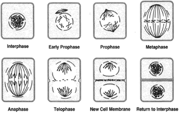 |
Figure 3-5 Plant cell mitosis. The steps are the same as in animal mitosis, but centrioles are not observed: interphase, early prophase, prophase, metaphase, anaphase, telophase, new cell wall beginning to form between daughter nuclei, and return to interphase |
produced from isolated carrot root cells. A proper word for this is totipotency. Any cell of a body has the capacity to produce a complete individual. This has been accomplished in the animal kingdom as well as in the plant kingdom. Frog intestine cells have been used successfully to produce frogs. This is done by enucleating an egg cell and then placing a nucleus from an intestine cell into the enucleated egg cell.
While the most common chromosome number of nonreproductive cells in plants is 2N sometimes 3N , 4N, and 5N do occur in such cells. The condition is called polyploidy and is further described later in this chapter.
Although one would expect the rate of mitosis to be a constant, experiments concerning frequency of mitoses in onion root tips suggest otherwise. Onions of uniform size were selected and provided with conditions that favored root growth, including constant light and temperature. Each hour of the day, a number of root tips were examined and the number of mitoses recorded. The results were as follows:
| Time of day: | 8 | 9 | 10 | 11 | 12 | 1 | 2 | 3 | 4 | |
|---|---|---|---|---|---|---|---|---|---|---|
| Number of mitoses: | 26 | 49 | 48 | 137 | 192 | 307 | 322 | 317 | 212 |
The preceding data show a maximum number of mitotic divisions at 2:OO P.M., when twelve times as many mitotic divisions occurred in an hour than were observed at 8:OO A.M. The frequency of mitoses, then, appears to be governed by circadian rhythms, or having a biological clock.
Support our developers


