In the Gymnophiona
In the Gymnophiona, the Saurobatrachia, and the Labyrinthodonta, the number of the vertebrae in the trunk is considerable, and the members of the two latter groups have long tails. But in the Batrachia, the total number of vertebrae does not exceed eleven, of which eight belong to the presacral region, one to the sacrum, and two (modified vertebrae) to the coccygeal region. The transverse processes of some of the presacral vertebrae are usually very long, but there are no separately ossified ribs. The transverse processes of the sacral vertebra are very large and expanded, and its centrum has usually a single concavity in front and a double convexity behind.The coccyx consists of a long, cylindroidal, basal bone proceeding from the ossification of the sheath of the termination of the notochord, and corresponding with the urostyle of the Teleostei; and of two neural arches, which lie over its anterior end, and become anchylosed with it. The anterior face of the coccyx usually presents two concave facets for articulation with the posterior convexities of the sacrum.
The cavity of the cranium is not narrowed anteriorly by the development of an interorbital septum in any Amphibian. All existing Amphibia have ex-occipitals developed in the walls of the cartilaginous cranium; but it is not certain that any such ossifications existed in Archegosaurus, though they are present in other Labyrinthodonts.
No Amphibian possesses a complete basi-occipital, supraoccipital, basisphenoid, alisphenoid, or presphenoid cartilage bone. In existing Amphibia, a prootic ossification appears to be very constant. The constant existence of distinct opisthotio and epiotic elements is doubtful.
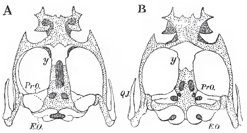 |
| Fig. 54. - The cartilagious cranium of Rana esculenta. A, from above; B, from below; y, the "os en ceinture," or girdle-bone. |
The membrane bones of the Amphibian skull are: 1. Frontals and parietals, which, in the Batrachia, may be fused together into one bone. 2. Nasals are generally present. 3. The vomers, always present, are two in number, one for each side, in all Amphibia but Pipa, Dactylethra, and Pelobates. 4. A great parasphenoid covers the base of the skull from the occipital to the ethmoidal region, as in Teleostei and Ganoidei. 5. A membrane bone (Z), called "temporo-mastoid" by Dugres, lies on the outer side of the suspensorium, extending from the side-walls of the skull to the articular head for the lower jaw. The relations of this bone in its upper part are similar to those of the squamosal of the higher Vertebrata, in its lower part to those of the bone F in Lepidosiren, to the preoperculum of fishes, and to the tympanic of the higher Vertebrata.
 |
| Fig. 55. - Skull of Rana esculenta. A, from above; B, from below; C. from the left side a, parasphenoid; y, girdle-bone; Z, the "temporo-mastoid" |
Two premaxillae are always developed. The maxillae are asually present, and may be connected, as in most Batrachia, by quadrato-jugal ossifications with the outer side of the end of the suspensorium, in which an ossification representing the quadrate bone is often developed. But the quadrato-jugals (and even the maxillae) may be represented simply by more or less ligamentous fibrous tissue, as is the case in the Urodela. Pterygoid bones are developed in all Amphibia, and distinct palatine bones in most, but not all, of the Batrachia. The suspensorium, which is inclined downward and forward in the lower Urodela, passes almost directly downward, or a little backward, in the higher, and in the Batrachia slopes greatly backward; and it undergoes the same modifications in direction, during the progress of any of the Batrachia from the larval to the adult state.
In the mandible, the proximal end of Meckel's cartilage is rarely, if ever, completely converted into a bony, articular element, but the distal moiety is ossified in some Batrachia. The membrane-bones of the mandible are a dentary and a splenial piece, with perhaps an angular element.
The hyoidean arch is, in most Amphibia, connected with the suspensorial cartilage - sometimes quite close to its origin, sometimes near its distal end, in the Urodela. Its cornua are stout and well ossified in the Proteidea. In the Batrachia they are slender, and their proximal ends may be free. Distally, they are connected with a broad lamellar body, from the posterior margin of which two processes which embrace the 'arynx are usually given off. In the perennibranchiate Proteidea, the hyoidean arches are united by narrow median entoglossal and urohyal pieces, as in Fishes.
In the Batrachia, the branchial arches disappear in the adult; but in the Gymnophiona and in the Urodela, more or fewer of the larval branchial arches persist throughout life.
Support our developers


