Cytogenetics
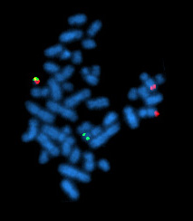
A metaphase cell positive for the bcr/abl rearrangement using FISH
History
Early years
Chromosomes were first observed in plant cells by Karl Wilhelm von N » geli in 1842. Their behavior in animal (salamander) cells was described by Walther Flemming, the discoverer of mitosis, in 1882. The name was coined by another German anatomist, von Waldeyer in 1888.The next stage took place after the development of genetics in the early 20th century, when it was appreciated that the set of chromosomes (the karyotype) was the carrier in the genes. Levitsky seems to have been the first to define the karyotype as the phenotypic appearance of the somatic chromosomes, in contrast to their genic contents. Investigation into the human karyotype took many years to settle the most basic question: how many chromosomes does a normal diploid human cell contain? In 1912, Hans von Winiwarter reported 47 chromosomes in spermatogonia and 48 in oogonia, concluding an XX/XO sex determination mechanism. Painter in 1922 was not certain whether the diploid number of man was 46 or 48, at first favouring 46. He revised his opinion later from 46 to 48, and he correctly insisted on man having an XX/XY system. Considering their techniques, these results were quite remarkable.
1. Using cells in culture
2. Pretreating cells in a hypotonic solution, which swells them and spreads the chromosomes
3. Arresting mitosis in metaphase by a solution of colchicine
4. Squashing the preparation on the slide forcing the chromosomes into a single plane
5. Cutting up a photomicrograph and arranging the result into an indisputable karyogram.
It took until the mid 1950s until it became generally accepted that the karyotype of man included only 46 chromosomes. Rather interestingly, the great apes have 48 chromosomes.
More Content on Cytogentics
» McClintock's work on maize
» Natural populations of Drosophila
» Human numerical abnormalities
» Advent of banding techniques
» Beginnings of molecular cytogenetics
» Medical uses
» Techniques
» Routine analysis
» Slide preparation
» Analysis
» Fluorescent in situ hybridization
» Slide preparation
» Analysis
» Future of cytogenetics
» References
Applications in biology
McClintock's work on maize
Barbara McClintock began her career as a maize cytogeneticist. In 1931 McClintock and Harriet Creighton demonstrated that cytological recombination of marked chromosomes correlated with recombination of genetic traits (genes). McClintock continued her career in cytogenetics studying the mechanics and inheritance of broken and ring (circular) chromosomes of maize. During her cytogenetic work, McClintock discovered transposons, a find which eventually led to her Nobel Prize in 1983.Natural populations of Drosophila
Evidence rapidly accumulated to show that natural selection was responsible. Using a method invented by L'Heretier and Teissier, Dobzhansky bred populations in population cages, which enabled feeding, breeding and sampling whilst preventing escape. This had the benefit of eliminating migration as a possible explanation of the results. Stocks containing inversions at a known initial frequency can be maintained in controlled conditions. It was found that the various chromosome types do not fluctuate at random, as they would if selectively neutral, but adjust to certain frequencies at which they become stabilised. By the time Dobzhansky published the third edition of his book in 1951 he was persuaded that the chromosome morphs were being maintained in the population by the selective advantage of the heterozygotes, as with most polymorphisms.
Human numerical abnormalities
Other numerical abnormalities discovered include sex chromosome abnormalities. An individual with only one sex chromosome (the X) has Turner syndrome, an additional X chromosome in a male, resulting in 47 total chromosomes, has Klinefelter's Syndrome. Many other sex chromosome combinations are compatible with live birth including XXX, XYY, and XXXX. The ability for mammals to tolerate aneuploidies in the sex chromosomes arises from the ability to inactivate them, which is required in normal females to compensate for having two copies of the chromosome. Not all genes on the X Chromosome are inactivated, which is why there is a phenotypic effect seen in individuals with an extra or missing X.
Trisomy 13 was associated with Patau's Syndrome and trisomy 18 with Edward's Syndrome.
Advent of banding techniques

Human male karyotype
Diagrams identifying the chromosomes based on the banding patterns are known as cytogenetic maps. These maps became the basis for both prenatal and oncological fields to quickly move cytogenetics into the clinical lab where karyotyping allowed scientists to look for chromosomal alterations. Techniques were expanded to allow for culture of free amniocytes recovered from amniotic fluid, and elongation techniques for all culture types that allow for higher resolution banding.
Beginnings of molecular cytogenetics
In the 1980s advances were made in molecular cytogenetics. While radioisotope-labeled probes had been hybridized with DNA since 1969, movement was now made in using fluorescently labeled probes. Hybridizing them to chromosomes preparations made using existing techniques came to be known as fluorescent in situ hybridization (FISH). This change significantly increased the usage of probing techniques as fluorescently labeled probes are safer and can be used almost indefinitely. Further advances in micromanipulation and examination of chromosomes led to the technique of chromosome microdissection whereby aberrations in chromosomal structure could be isolated, cloned and studied in ever greater detail.Medical uses

translocation 9;11 associated with AML
In congenital disorders, such as Down's syndrome, cytogenetics can determine the nature of the chromosomal defect - a "simple" trisomy, a mosaic, "balanced" translocation, a deletion, or an insertion in one - or both - of the parents, or in the fetus. With the advent of harvest procedures which allowed easy enumeration of chromosomes, discoveries were quickly made in abnormalities arising from nondysjunction events which cause cells with aneusomy (additions or deletions of entire chromosomes). In 1959 Lejeune discovered patients with Down syndrome had an extra copy of chromosome 21. Down syndrome is also referred to as trisomy 21. In 1960 Nowell discovered a small chromosome, dubbed the Philadelphia chromosome, which was shown to be the cause of Chronic myelogenous leukemia. 13 years later this was shown by Janet Rowley to be a translocation of chromosomes 9 and 22.
Many other sex chromosome combinations are compatible with live birth including XXX, XYY, and XXXX. The ability for mammals to tolerate aneusomies in the sex chromosomes arises from the ability to inactivate them, which is required in normal females to compensate for having two copies of the chromosome.
Trisomy 13 was associated with Patau's Syndrome and trisomy 18 with Edward's Syndrome.
Techniques
Routine analysis
Several chromosome-banding techniques are used in cytogenetics laboratories. Quinacrine banding (Q-banding) was the first staining method used to produce specific banding patterns. This method requires a fluorescence microscope and is no longer as widely used as Giemsa banding (G-banding). Reverse banding (R-banding) requires heat treatment and reverses the usual white and black pattern that is seen in G-bands and Q-bands. This method is particularly helpful for staining the distal ends of chromosomes. Other staining techniques include C-banding and nucleolar organizing region stains (NOR stains). These latter methods specifically stain certain portions of the chromosome. C-banding stains the constitutive heterochromatin, which usually lies near the centromere, and NOR staining highlights the satellites and stalks of acrocentric chromosomes. High-resolution banding involves the staining of chromosomes during prophase or early metaphase (prometaphase), before they reach maximal condensation. Because prophase and prometaphase chromosomes are more extended than metaphase chromosomes, the number of bands observable for all chromosomes increases from about 300 to 450 to as many as 800. This allows the detection of less obvious abnormalities usually not seen with conventional banding.
Slide preparation
Analysis
Analysis of banded chromosomes is done at a microscope by a clinical laboratory specialist in cytogenetics (CLSp(CG)). Generally 20 cells are analyzed which is enough to rule out mosaicism to an acceptable level. The results are summarized and given to a board-certified medical geneticist and a pathologist for review, and to write an interpretation taking into account the patients previous history and other clinical findings. The results are then given out reported in an International System for Human Cytogenetic Nomenclature 2005 (ISCN2005).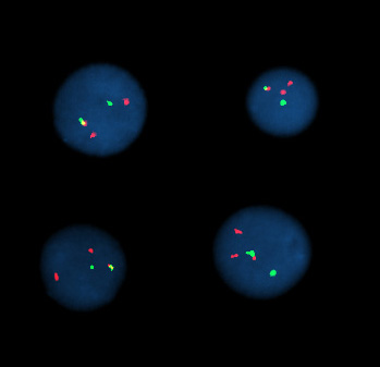
Interphase cells positive for a t(9;22) rearrangement
Fluorescent in situ hybridization
Fluorescent in situ hybridization refers to using fluorescently labeled probe to hybridize to cytogenetic cell preparations.In addition to standard preparations FISH can also be performed on:
» bone marrow smears
» blood smears
» paraffin embedded tissue preparations
» enzymatically dissociated tissue samples
» uncultured bone marrow
» uncultured amniocytes
» cytospin preparations
Slide preparation
This section refers to preparation of standard cytogenetic preparationsThe slide is aged using a salt solution usually consisting of 2X SSC (salt, sodium citrate). The slides are then dehydrated in ethanol, and the probe mixture is added. The sample DNA and the probe DNA are then co-denatured using a heated plate and allowed to re-anneal for at least 4 hours. The slides are then washed to remove excess unbound probe, and counterstained with 4',6-Diamidino-2-phenylindole (DAPI) or propidium iodide.
Analysis
Analysis of FISH specimens is done by fluorescence microscopy by a clinical laboratory specialist in cytogenetics (CLSp(CG)). For oncology generally a large number of interphase cells are scored in order to rule out low level residual disease, generally between 200 and 1000 cells are counted and scored. For congenital problems usually 20 metaphase cells are scored.Future of cytogenetics
Some References on Cytogentics
-
Stedman's Medical Dictionary (28th Ed.). (2006). Baltimore, MD: Lippincott Williams.
-
Levitsky G.A. 1924. The material basis of heredity. State Publication Office of the Ukraine, Kiev. [in Russian]
-
Levitsky G.A. 1931. The morphology of chromosomes. Bull. Applied Bot. Genet. Plant Breed. 27, 19-174.
-
Kottler M. 1974. From 48 to 46: cytological technique, preconception and the counting of the human chromosomes. Bull. Hist. Med. 48, 465-502.
-
von Winiwarter H. 1912. Études sur la spermatogenese humaine. Arch. biologie 27, 93, 147-9.
-
Painter T.S. 1922. The spermatogenesis of man. Anat. Res. 23, 129.
-
Painter T.S. 1923. Studies in mammalian spermatogenesis II. The spermatogenesis of man. J. Exp. Zoology 37, 291-336.
-
Tjio J.H & Levan A. 1956. The chromosome number of man. Hereditas 42, 1-6.
-
Hsu T.C. Human and mammalian cytogenetics: a historical perspective. Springer-Verlag, N.Y.
-
Painter T.S. 1933. A new method for the study of chromosome rearrangements and the plotting of chromosome maps. Science 78: 585-586.
-
Dobzhansky T. 1951. Genetics and the origin of species. 3rd ed, Columbia University Press, New York.
-
Dobzhansky T. 1970. Genetics of the evolutionary process. Columbia University Press N.Y.
-
[Dobzhansky T.] 1981. Dobzhansky's genetics of natural populations. eds Lewontin RC, Moore JA, Provine WB and Wallace B. Columbia University Press N.Y.
-
Lejeune J, Gautier M, Turpin MR. Etude des chromosomes somatiques de neuf enfants mongoliens. C R Acad Sci (Paris) 1959;248:1721-2.
- Nowell PC, Hungerford DA. A minute chromosome in human chronic granulocytic leukemia. Science 1960;132:1497-1501.
Support our developers


