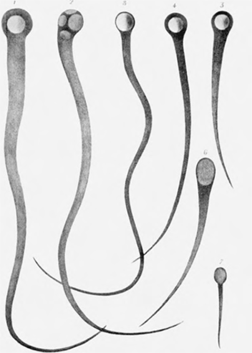
Figure 1-18 An early nineteenth-century micrographic drawing of sperm from (1) guinea pig, (2) white mouse, (3) hedgehog, (4) horse, (5) cat, (6) ram, and (7) dog (Prévost and Dumas, 1821). Some biologists initially interpreted these as parasitic worms in the semen, but on further examination found them to be male gametes.

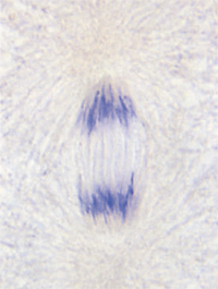
Figure 1-19 Paired chromosomes are physically associated and then segregated into different daughter cells during cell division prior to gamete formation
Improvements in microscopes during the 1800s permitted cytologists to study the production of gametes by direct observation of reproductive tissues. Interpreting the observations was initially difficult, however. Some prominent biologists hypothesized, for example, that sperm were parasitic worms in the semen (Figure 1-18). This hypothesis was soon falsified, and the true nature of gametes was clarified. As the precursors of gametes prepare to divide in the early stages of gamete production, the nuclear material condenses to reveal discrete, elongate structures called chromosomes. Chromosomes occur in pairs that are usually similar but not identical in appearance and informational content. The number of chromosomal pairs varies among species. One member of each pair is derived from the female parent and the other from the male parent. Paired chromosomes are physically associated and then segregated into different daughter cells during cell division prior to gamete formation (Figure 1-19). Each resulting gamete receives one chromosome from each pair. Different pairs of chromosomes are sorted into gametes independently of each other. Because the behavior of the chromosomal material during gamete formation parallels that postulated for Mendel's genes, Sutton and Boveri in 1903 through 1904 hypothesized that chromosomes were the physical bearers of the genetic material. This hypothesis met with extreme skepticism when first proposed. A long series of tests designed to falsify it nonetheless showed that its predictions were upheld. The chromosomal theory of inheritance is now well established.

Figure 1-18 An early nineteenth-century micrographic drawing of sperm from (1) guinea pig, (2) white mouse, (3) hedgehog, (4) horse, (5) cat, (6) ram, and (7) dog (Prévost and Dumas, 1821). Some biologists initially interpreted these as parasitic worms in the semen, but on further examination found them to be male gametes.

Figure 1-19 Paired chromosomes are physically associated and then segregated into different daughter cells during cell division prior to gamete formation


