Motility in the Alimentary Canal
 |
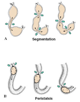
| Figure 34-8 Movement of intestinal contents by segmentation and peristalsis. A, Segmentational movements of food showing how constrictions squeeze the food back and forth, mixing it with enzymes. The sequential mixing movements occur at about 1-second intervals. B, Peristaltic movement, showing how food is propelled forward by a traveling wave of contraction. |
Motility in the Alimentary Canal
Food is moved through the digestive tract by cilia or by specialized musculature, and often by both. Movement is usually by cilia in the acoelomate and pseudocoelomate metazoa that lack the mesodermally derived gut musculature of true coelomates. Cilia move intestinal fluids and materials also in some eucoelomates, such as most molluscs, in which the coelom is weakly developed. In animals with well-developed coeloms, the gut is usually lined with two opposing layers of smooth muscle: a longitudinal layer, in which the smooth muscle fibers run parallel with the length of the gut, and a circular layer, in which the muscle fibers embrace the circumference of the gut. The most characteristic gut movement is segmentation, the alternate constriction of rings of smooth muscle of the intestine that constantly divide and squeeze the contents back and forth (Figure 34-8A). Walter B. Cannon of homeostasis fame, while still a medical student at Harvard in 1900, was the first to use X rays to watch segmentation in experimental animals that had been fed suspensions of barium sulfate. Segmentation serves to mix food but does not move it through the gut. Another kind of muscular action, called peristalsis, sweeps the food down the gut with waves of contraction of circular muscle (Figure 34-8B).


