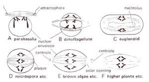
Fig. 7.2. Mitotic anaphase in some organisms, showing variation in the disappearance of nuclear membrane.

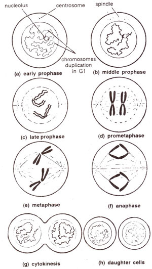
Fig. 7.3. Various stages of mitosis in a somatic cell.
At prophase (after DNA synthesis is already completed during interphase), the cell is still preparing for division. At the beginning of prophase chromosomes appear as thin, filamentous uncoiled structures (Fig. 7.3a). Chromosomes become coiled, shortened and more distinct as the mitosis progresses through prophase, which is of much longer duration than other stages (Fig. 7.3b, c, d). The durations of different stages of mitosis in four organisms are presented in Table 7.2.
An important characteristic of mitotic prophase is longitudinal splitting of each chromosome into two sister chromatids (Fig. 7.3c, 7.3d). Double structure of each chromosome thus may be conspicuous at late prophase. This feature can be easily observed in suitably fixed plant or animal chromosomes. Sister chromatids at this stage are attached only at
centromere and it will be at this position that two chromatids will now remain attached to spindle tubule. Soon after the nuclear membrane disappears at late prophase (Fig. 7.3c). Similarly, nucleolus also disappears before the cell enters mitotic metaphase. However, there are variations available with respect to the disappearance of nuclear membrane and the nucleolus. In a number of primitive plant and animal species, the nuclear membrane does not dissolve. Some of these variations are shown in Figure 7.2.

Fig. 7.2. Mitotic anaphase in some organisms, showing variation in the disappearance of nuclear membrane.

Fig. 7.3. Various stages of mitosis in a somatic cell.


