Cellular Organization
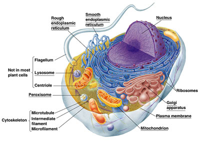
Structurally, there are two basic kinds of cells: prokaryotic and eukaryotic. Prokaryotic cells, including bacteria and archae, although far from simple, are generally much smaller and less complex structurally than eukaryotic cells. The major difference is that the genetic material (DNA) is not sequestered within a double-membrane structure called a nucleus (Figure 1-1). In eukaryotes, a complete set of genetic instructions is found on the DNA molecules, which exist as multiple linear structures called chromosomes that are confined within the nucleus.Eukaryotic cells also contain other membrane-bound organelles within their cytoplasm (the region between the nucleus and the plasma membrane). These subcellular structures vary tremendously in structure and function.
Most eukaryotic cells have mitochondria, which contain the enzymes and machinery for aerobic respiration and oxidative phosphorylation. Thus, their main function is generation of adenosine triphosphate (ATP), the primary currency of energy exchanges within the cell. This organelle is bounded by a double membrane. The inner membrane, which houses the electron transport chain and the enzymes necessary for ATP synthesis, has numerous foldings called cristae, which protrude into the matrix, or central space. Mitochondria contain their own DNAand ribosomes, but most of their proteins are imported from the cytoplasm.
Chloroplasts contain the photosynthetic systems for utilizing the radiant energy of sunlight and are found only in plants and algae. Photosynthesis is the process that converts light energy into the chemical bond energy of ATP, which in turn can be used to convert carbon dioxide (CO2) and water (H2O) into carbohydrates. Chloroplasts contain an internal system of membranes called thylakoids, a circular chromosome, and their own ribosomes. The flattened, vesicular thylakoids contain the chlorophyll pigments, the enzymes, and other molecules needed to harness light energy for conversion to chemical energy. Carbon fixation occurs in the stroma, the space between the thylakoids and the inner membrane.
 |
| Figure 1-1 An animal cell. |
Prokaryotic cells lack internal membranes, but photosynthetic bacteria contain invaginations of the plasma membrane called mesosomes.
Centrioles, located within the centrosome, are associated with the cell’s polar regions, toward which the chromosomes migrate during cell division, and are found only in animal cells. The endoplasmic reticulum (ER) amplifies the surface area available for specialized biochemical reactions and the synthesis of certain types of proteins. The Golgi complex directs the transport of proteins and other biomolecules to specific locations within the cell. Vacuoles serve as storage compartments for food, water, or other molecules. Enzymes digest materials brought into the cell within lysosomes.
Ribosomes function in the manufacture of proteins. The ribosomes in prokaryotes are smaller than those found in the cytoplasm in eukaryotes, but are similar in size and structure to those found in the mitochondria and chloroplasts of eukaryotes. Eukaryotic ribosomes associated with the ER give it a granular appearance, hence the name rough ER.
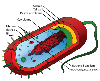
Motility is accomplished by different means in prokaryotic and eukaryotic cells. Eukaryotic cells, such as amoebas and white blood cells, creep along substrates as an undulating mass of constantly changing morphology. This type of motion is achieved by a massive network of protein fibers, the cytoskeleton. Motile bacteria are usually propelled by one or more hairlike appendages called flagella that originate in the plasma membrane and rotate like propellar shafts (Figure 1-2). These filaments are constructed of the protein flagellin. Some eukaryotic cells also have flagella, but they consist of bundles of microtubules made of tubulin, and they originate from a basal body in the cytoplasm. Eukaryotic flagella such as those in sperm tails bend back and forth in quasi-sinusoidal waves. Eukaryotic cilia are structurally similar but are much shorter, more numerous, and more rigid on the powerstroke. Some bacteria also have long hollow tubes called pili or fimbriae composed of a protein called pilin. These structures do not contribute to motility, but to the adhesiveness of bacteria and the facilitation of conjugation.
One of the distinguishing features between plants and animals is that plants and fungi have cell walls made of cellulose and chitin, respectively, but animal cells do not. Almost all bacteria have a rigid cell wall surrounding the plasma membrane, but it has a different structure than the plant cell wall and is composed of peptidoglycan. Some bacteria also have a polysaccharide capsule or a glycocalyx surrounding the cell wall.
 |
| Figure 1-2 A bacterial cell. |
These protect the bacteria from predatory cells and promote their attachment to various objects and to each other. Most eukaryotic cells also have a glycocalyx that covers the surface of the cell and promotes cell adhesions in the formation of specific tissues. In addition, many types of animal cells are surrounded by an extracellular matrix, which comprises a variety of proteins that give specific tissues their characteristic properties.
Notes:
Mitochondria are nicknamed the “powerhouses” of the cell because of their role in ATP production.
Proteins that are:
1. membrane bound
2. secreted
3. compartmentalized
are synthesized on the ER.


