Deficiency Symptoms
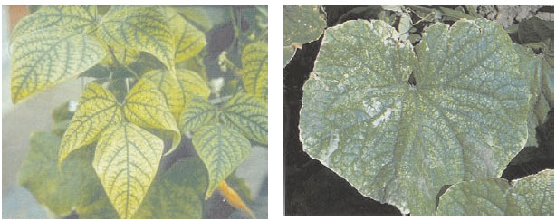
Characteristic foliar symptoms of manganese deficiency become unmistakable only when the growth rate is restricted significantly (67) and include diffuse interveinal chlorosis on young expanded leaf blades (Figure 12.1) (60); in contrast to the network of green veins seen with iron deficiency (67). Severe necrotic spots or streaks may also form. Symptoms often occur first on the middle leaves, in contrast to the symptoms of magnesium deficiency, which appear on older leaves. With eucalyptus (Eucalyptus spp. L. Her.), the tip margins of juvenile and adult expanding leaves become pale green. Chlorosis extends between the lateral veins toward the midrib (60). With cereals, chlorosis develops first on the leaf base, while with dicotyledons the distal portions of the leaf blade are affected first (67). |
| FIGURE 12.1 Manganese deficiency on crops: left: garden bean (Phaseolus vulgaris L.) and right, cucumber (Cucumis sativus L.). |
With citrus, dark-green bands form along the midrib and main veins, with lighter green areas between the bands. In mild cases the symptoms appear on young leaves and disappear as the leaf matures. Young leaves often show a network of green veins in a lighter green background, closely resembling iron chlorosis (75). Manganese deficiency is confirmed by the presence of discoloration (marsh spot) on pea seed cotyledons (87), and split or malformed seed of lupins (84).
In contrast to iron deficiency chlorosis, chlorosis induced by manganese deficiency is not uniformly distributed over the entire leaf blade and tissue may become rapidly necrotic (88). The inability of manganese to be re-translocated from the old leaves to the younger ones designates the youngest leaves as the most useful for further chemical analysis to confirm manganese deficiency. Visual symptoms of manganese deficiency can easily be mistaken for those of other nutrients such as iron, magnesium, and sulfur (87), and vary between crops. However, they are a valuable basis for the determination of nutrient imbalance (87) and, combined with chemical analysis, can lead to a correct diagnosis.


