The Cetacea
In this order of Mammalia the form of the body is still more fish-like than in the Sirenia. There is no trace of a neck, the contour of the head passing gradually into that of the body. A horizontally-flattened caudal fin is always present; and, very generally, the dorsal integument is produced into a median, laterally-compressed dorsal fin. The body is incased in a thick smooth integument, beneath which a very thick layer of fat is deposited. Hairs are almost entirely absent in the adult state.As in the recent Sirenia, the anterior limbs alone are present. Externally they do not present any indication of division into brachium, antibrachium, and manus, but have the form of a broad, flattened paddle, without any vestiges of nails.
The one or two apertures by which the cavity of the nose opens externally, are always situated at the top of the head, and far removed from the extremity of the snout. There is no third eyelid, and the very small auditory apertures are totally devoid of any pinna. The teats are two, and, in the female, are lodged in depressions on each side of the vulva.
The articular surfaces of the centra of the vertebrae are flat, and the epiphyses usually remain distinct for a long time.
The spinal column, as a whole, is remarkable for the shortness of its cervical, and the length of its lumbar region, there being sometimes a greater number of lumbar than of dorsal vertebrae. There is no sacrum. The caudal vertebrae are only distinguishable from the posterior lumbo-sacral vertebrae by their chevron-bones. The second vertebra of the neck is devoid of any odontoid process; and it very commonly happens that more or fewer of the cervical vertebrae, the bodies of which are often so short as to be mere disks, are anchylosed together, either by their arches, or by their centra, or by both. The centra of all the succeeding vertebrae are large in proportion to their arches, and the inter-vertebral fibro-cartilages are exceedingly thick, so as to confer great flexibility and elasticity on the spine. The arches of the hinder dorsal vertebrae, and of those of the lumbar and caudal regions, are not articulated together by zygapophyses. The centra of the posterior caudal vertebras lose their processes and become polygonal.
Very few of the ribs become connected with the sternum at their distal ends; and, in contradistinction to what happens in most Mammalia, the proximal ends of the majority of the ribs are connected only with the transverse processes of the vertebrae, and not with their bodies.
The skull is even more remarkably modified than the vertebral column. The brain-case itself has a spheroidal form; while the jaws are greatly prolonged, the principal enlargement of the upper jaw taking place in the region which lies in front of the nasal aperture. The basis cranii, as a whole, is ramarkably broad, and its upper surface concave from before backward, the sella turcica being very slightly indicated. The parietal bones are comparatively small, and do not meet in a sagittal suture, as they do in other Mammalia; the supraoccipital, with an interparietal bone, being interposed between them, and extending forward so as to unite with the frontals. Each frontal bone is produced outward into a great bony plate which covers the orbit. The squamosal bone sends a very large and stout zygomatic process forward to meet this supra-orbital prolongation of the frontal. The proper jugal bone, on the other hand, which bounds the orbit below, is exceedingly slender. The very large maxilla extends backward and outward in contact with the frontal, or even overlapping the greater part of its surface; and it stretches forward to very near the anterior end of the snout, so that almost the whole of the gape is bounded by the maxilla.
The premaxillae, on the other hand, though very long, inasmuch as they occupy the whole length of the jaw in the middle line, from the anterior nasal aperture to the end of the snout, are almost entirely excluded from the gape.
The nasal bones are always short; and, sometimes, are mere bony tuberosities united with the frontal bones behind the anterior nasal aperture. The turbinal bones are almost always rudimentary, and the nasal passages are nearly vertical, in consequence, for the most part, of the rudimentary condition and shortness of the nasal bones.
The periotic bones are loosely connected with the squamosal and tympanic, and are usually united with the other bones of the skull only by cartilage, so that they fall out very readily in the dry skull. The tympanic bones are commonly of very considerable size, thick and scroll-shaped.
The lower jaw has hardly any coronoid process, and its ramus has no perpendicular portion, the condyle being situated upon its posterior extremity. The body of the hyoid is a very broad plate of bone, and has two pair of stout, well-ossified cornua.
The Cetacea are devoid of clavicles. If the spine of the scapula is present, it is a low ridge situated close to the anterior edge of the bone; but it commonly terminates in a long acromion process, and, sometimes, there is a conspicuous, straight, and flattened coracoid. The humerus is short, and the articular surfaces at its distal end are, in all recent Cetacea, flat facets inclined to one another at an angle. The ulna and the radius are short, laterally-compressed bones, without any movement upon one another; and, in all recent Cetacea, they are not freely movable upon the humerus. The carpus is often imperfectly ossified. When the carpal bones are complete, they are polygonal and imbedded in a fibrous tissue; not united by articulations provided with synovial membranes. The digits do not exceed five in number, but there are always more than three phalanges in some of them.
The pelvis is represented by two bones which lie parallel with the axis of the vertebral column, give attachment to the corpora cavernosa in the male, and, therefore probably represent the ischia. They are elongated, convex upward and concave downward, and are connected with the vertebral column only by fibrous tissue. In some few Cetacea (Balaenoidea) ossicles, which lie on the outer side of the pelvic bone, appear to represent the femur, but no further indication of a hindlimb has been discovered.
In most of the Cetacea, the muscles which, in other Mammalia, move the antibrachium and the manus, are absent, those which move the humerus upon the shoulder-blade being, alone, represented.
In no recent Cetacean have the teeth any vertical successors, nor more than a single root. The alveoli are often incompletely separated from one another. The number of the teeth varies very greatly, but they are almost alwavs nearly uniform in character. There appear to be no salivary glands. The stomach is complicated, being divided into, at fewest, three chambers, of which the first is a kind of paunch lined by a thick epithelium, while the second and the third are more elongated, the last stomach being that in which digestion takes place.
The arteries and veins form great plexuses, or relia mirabila, and these are especially conspicuous in the cavity of the thorax, upon each side of the vertebral column, and in the intercostal spaces.
The soft palate is remarkably long and muscular. The epiglottis and the arytenoid cartilages are more or less produced, so as to give the glottis the shape of a funnel, the apex of which is embraced by the soft palate, in such a manner as to form a continuous air-passage from the posterior nares to the larynx, on each side of which the food passes. The very short trachea, before it divides into the bronchi, gives off the so-called "third bronchus" to the right lung, as in the Bears, Walruses, and Ruminants.
 |
| Fig. 104. - Lateral and superior views of the skull of a foetal Whale (Balaena Australia)-The jugal bones are absent, and the figure does not sufficiently indicate the outward curvature of the ramus of the mandible (Mn.). |
The Cetacea are divisible into three groups: the Balaenoidea, the Delphinoidea, and the Phocodontia (For further information respecting the characters of the recent Cetacea, I refer the reader to Prof. Flowers's very valuable memoir "On the Osteology of Inia and Fontoporia," published in the "Transactions of the Zoological Society for 1867.")
a. In the Balaenoidea the nasal chambers communicate with the exterior by two apertures, which are capable of being shut at the will of the animal, and are called spiracles. These are not connected with any saccular dilatations of the nasal passages, situated between the skull and the integument.
In the spinal column, no rib has a complete neck and capitulum, the heads of even the most anterior ribs being united with the bodies of the vertebrae only by ligament. The chief connection of all the ribs, therefore, and the only connection of most of them, is with the transverse processes of the vertebrae. The short and broad sternum unites only with the first rib, and the union is direct, so that there are no sternocostal libs.
The skull (Fig. 104) is exceedingly large in proportion to the body, and nearly symmetrical. The nasal bones, Nu., though short, are longer, and more like those of ordinary mammals, than is the case in other Cetacea. The maxilla, Mx., extends outward in front of the great supraorbital process of the frontal, Fr., but it does not cover the frontal bone. There is a distinct lachrymal. Each ramus of the mandible, Mn., is convex outward and concave inward; and the space between the rami of the mandible is very much greater than the width of the maxillo-premaxillary part of the skull, which tapers to its anterior end, and is more or less convex upward and concave inferiorly. The two rami of the mandible are connected only by ligament at the symphysis.
 |
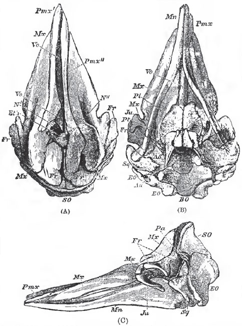
| Fig. 105. - "Ear-bones" of the adult Balaena Australia. - Seen from within in the upper figure; from without in the lowor. Eu., Eustachian canal; Au.,external auditory meatus; Sty., ossified root of the styloid process. |
In some of the Balaenoidea, e.g., Balaena rostrata, the cricoid cartilage and the rings of the trachea are incomplete in front, and a large air-sac is developed in the cricothyroid space. The Balaenoidea possess olfactory nerves and a distinct, though small, olfactory apparatus. The sclerotic coat of the eyeball is enormously thick, and the optic nerve is surrounded by a rete-mirdbile. The tympanic membrane is connected with the malleus by ligament. The semicircular canals are very small, but the cochlea is large, and makes only 1 1/2 turns. The muscles of the antibrachium and manus are not altogether absent.
 |
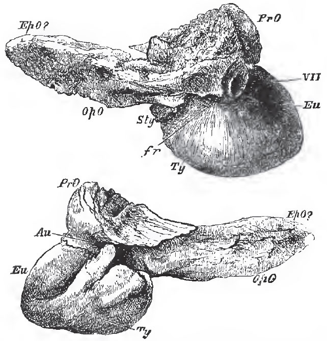
| Fig. 106. - Upper (A), under (B), and lateral (C) views of the skull of a foetal Cachalot (Physeter).-The nasal bones arc not presented in the upper view, and the hinder ead of the jugal is displaced from its natural connection with the squamosal in (C), |
b. In the Delphinoidea the nasal chambers open by only a single spiracle on the top of the head; and saccular dilatations of various dimensions are developed from the walls of the passage which connects this aperture with the bony nasopalatine passages, and lie between the integument and the outer surface of the skull.
More or fewer of the anterior ribs have heads and necks, the capitula articulating with the bodies of the vertebrae, as in other Mammalia. The elongated sternum is, almost always, composed of several pieces arranged in a longitudinal series; and cartilaginous, or ossified, sternal ribs are present in greater or smaller number. The nasal bones, which are very short, and have their upper surfaces tubercle-like, are more or less asymmetrically developed, as are also the maxillae; so that the facial part of the skull appears distorted. The maxillae are expanded behind, and cover the orbital process of the frontal bone wholly or partially. The lachrymal bone is usually small and confluent with the slender jugal, but it may be large and distinct. The rami of the mandible are not arcuated outward, and they become united in a longer or shorter symphysis. The mandible, as a whole, is not sensibly broader than the corresponding portion of the maxillo-premaxillary part of the skull.
Teeth always exist after birth, and are never replaced by baleen plates. They are usually numerous, but sometimes few and deciduous. Occasionally, only one or two teeth persist, and these, as in the Narwhal, may take the form of immensely- elongated tusks.
To this division belong the Physeteridae Platanistidae and Delphinidae.
The Physeteridae possess functional teeth only in the lower jaw. The asymmetry of the skull is strongly pronounced; and, in the adult, the maxillary and frontal bones are produced, so as to form a sort of basin upon the upper and anterior surface of the skull. The pterygoids meet in the middle line below, and the mandibular symphysis is sometimes extremely long.
The greater number of the cervical vertebrae are anchylosed. The hinder ribs lose their tubercular, but retain their capitular articulation with the vertebrae. The costal cartilages are not ossified. The pectoral limbs are small, and a dorsal fin is usually present.
The proper Sperm Whales (Physeterinae) have an enormous head, with a quadrate truncated snout, at the anterior superior angle of which the spiracle is placed. The teeth become fully developed only in the lower jaw. The cranial basin is immense, and is filled by a loose connective tissue, in which the peculiar fat known as spermaceti is contained. Ambergris is a sort of bezoar, found in the alimentary canal of tho Cachalot, and seemingly derived from the fatty matter contained in the Cephalopoda on which the Cetacean feeds. In the other group of the Physeteridae,-the Ziphiinae or Rhynchoceti- to which the Bottlenosed Whale (Hyperoodon) belongs, there are only one or two pairs of fully-formed teeth in the mandible. Some recent and many fossil (middle and later tertiary) genera of the Cetaceans are remarkable for the elongated rostrum formed by the solid ossification and anchylosis of the ethmoid, premaxillae, and maxillae.
The Platanistidae are fluviatile or estuarine Cetacea, which occur in the Ganges and in the rivers of South America. The cervical vertebrae are not anchylosed, and the costal cartilages are not ossified. The tubercula and capitula of the ribs blend together posteriorly. The symphysis of the mandibles is extremely long and the jaws are narrow. Numerous teeth with compressed fangs are found in both jaws. The eyes are small, and in Platanista they are rudimentary.
In the Dephinidae lastly (Dolphins, Porpoises, Grampuses), the teeth are usually numerous in both jaws, though the Narwhal is an exception to this rule, as has already been mentioned.
The anterior cervical vertebrae are generally anchylosed together. The posterior ribs lose their capitula and become articulated only with the transverse processes of the vertebrae. The costal cartilages are well ossified. The symphysis of the mandible does not exceed one third of the rami in length, and the frontal and maxillary bones are not especially produced upward at their edges.
As the common Porpoise (Phoccaena communis), which is a member of this group, is the Cetacean which is most likely to come within reach of the student, it may be useful to speak at some length of its more interesting anatomical peculiarities.
The adult animal is usually about five feet long, and is covered with a smooth integument upon which no hair is to be discovered, though a few hairs are visible about the mouth in the young animal. The contour of the anterior part of the head is very convex, and presents, in the middle line, the spiracle or blow-hole, which has the form of a crescent with the points turned downward and forward. The eyes are small end placed low down, close to the posterior end of the gape of the mouth, which is bounded by dense and rigid lips. The aperture of the ear lies about an inch and three-quarters behind the eye, and is so minute as to be discovered with difficulty. The genital aperture is placed a long way in front of the anus in the male; while, in the female, the interval, in which the fossse which lodge the teats are situated, is much less. There is a conspieuous vertical dorsal fin in addition to the flattened caudal fin. Immediately beneath the skin is a thick layer of blubber, as in other Cetacea.
In the spongy texture of all the bones, the absence of medullary cavities in those of the limbs, and in the long persistent separability of the epiphyses of the centra of the vertebrae, the Porpoise resembles other Cetacea; as it does in the shortness of the cervical, and the length of the lumbar, region of the spinal column.
The seven cervical vertebrae are all anchylosed together, and the atlas, which is very large in proportion to the rest, overlaps them above and at the sides. The centra of the hinder cervicals are so short and broad that they are mere plates of bone. There are twenty-eight dorso-lumbar vertebrae, of which fifteen are dorsal. In all but the most anterior of these vertebrae, the zygapophyses are abortive; and long accessory processes, developed from the front-part of the neural arches, loosely embrace the spine of the vertebrae in front. This arrangement, together with the thickness of the intervertebral ligaments, gives great flexibility to the spinal column. The transverse processes of the hinder dorsal, and of the lumbar, vertebra are very long. There are five pairs of true ribs. The sternebrae anchylose into an elongated sternum. The anterior caudal vertebrae are provided with large chevron-bones, and their transverse processes exhibit notches through which branches of the aorta pass.
In consequence of the globular form of the brain-case, and the prolongation of the jaws, the skull has a flask-like shape. There is a slight want of symmetry about the base of the upper jaw, but it is hardly appreciable.
In a longitudinal section, the flatness and the upwardly concave contour of the base of the skull; the extreme shallowness of the sella turcica; the presence of an ossified tentorium; and the broad imperforate anterior wall, in the place of the cribriform plate of the ethmoid, are striking features. The synchondrosis between the basi-and presphenoid is persistent. On the base of the skull the basi-occipital gives off great processes outward and downward, to form, together with a paramastoid prolongation of the exocoipital, and the squamosal, a chamber in which the anchylosed tympanie and periotic bones are contained. The ex-and supra-occipitals, together with the interparietals, form the whole back wall and middle of the roof of the cranium, separating the parietals completely, and the frontals largely, and reaching the nasal bones.
The basi-sphenoid is anchylosed with the small and almost horizontal alisphenoids, and there are no sphenoidal pterygoid processes. The parietals are small, and occupy only the under and lateral portions of the brain-case. The frontal bones are very broad and expanded, and are completely anchylosed together, where they form the front wall of the brain-case. Posteriorly and above, they diverge to receive the interparietal. The supra-orbital processes are extremely large, and are directed forward and outward, not backward and outward, as in the Whalebone Whales. The greater part of the superior surface of the frontals and of their orbital processes is rough and covered over by the expanded maxillary bones, which allow only a narrow, transverse, smooth, band-like surface, formed by the frontals, to be seen on the upper and anterior region of the skull. The rough surface is marked by two shallow grooves which pass from below upward, and are convex toward one another and to the middle line. Corresponding grooves exist on the under side of the expanded proximal ends of the maxillaries; and, when these are in their natural positions, the coadapted grooves form two canals, which are blind in front and above. These, in the natural state, are full of air, and communicate with the air-chambers at the base of the skull and with the Eustachian tubes.
The narrow premaxillae are anchylosed with the inner margins of the maxillae, and contribute only a very small portion of the alveolar margin of the upper jaw. The alveoli are not completely separated from one another. The pterygoid bones do not unite in the palate. They have a peculiar excavated form, and are notched for the passage of the ends of the Eustachian tubes into the nasal passages. These are nearly vertical and are separated by the large and strong vomer. Their superior apertures are left quite uncovered in consequence of the small size, tubercular form, and backward position of the nasal bones. The squamosal is relatively small, but has the characteristically cetacean, large, zygomatic process; this extends forward nearly to the posterior end of the supraorbital process, and gives attachment to the slender jugal.
The periotic bones form a dense osseous mass, which is anchylosed with the no less heavy and thick, scroll-shaped tympanic. The pars mastoidea of the periotic mass fits pretty accurately into a recess of the chamber which has already been described; and is thus held in position in the dry skull, though it is very easily detached.
When the tympano-periotic bone and all the facial bones are removed, only two pair of foramina are visible in the base of the skull. The anterior pair give exit to the second, third, fourth, the anterior division of the fifth and the sixth nerves, and these answer to the optic and sphenorbital foramina. The posterior pair take the place of the oval, posterior lacerated, and jugular foramina, and the precondyloid foramina open into them posteriorly. The rami of the mandibles are only united by a short symphysis. The body of the hyoid is broad and hexagonal, and has two slender, anterior, and two broad and flat, posterior, cornua.
In the natural position the fore-limbs stand out from the body with their flat surfaces looking upward and downward; the upper surface being directed a little backward, and the lower a little forward. The tuberosity of the short humerus is directed forward. The carpus contains six or seven ossifications. The number of phalanges in the digits is two, eight, six, three, two, counting the pollex as the first.
The pelvic bones are elongated, slightly curved, osseous styles. They lie with their long axes parallel to the vertebral column, their convex sides upward, and their smaller ends forward, within an inch of the centra of the vertebrae, their hinder-ends being close to the third chevron-bone of the tail. The front-ends are about an inch apart. Behind its centre, each bone presents a flattened thickening for the attachment of the corpus cavemosum of its side.
The cutaneous muscle is very largely developed, and lies between two layers of blubber, the thick surperficial one separating it from the skin, and the thin deep layer from the subjacent muscles. It may be said to be disposed in two broad layers, a dorsal and a ventral, on each side; these extend from the occipital crest, and from the rami of the mandibles, to the tail. Both these divisions send off strong bundles to the humerus, which act as powerful adductors, abductors, protractors, and retractors of the fin. There is no trapezius, and the representative of the latissimus dorsi is very small. A strong occipito-humeralis, from the paramastoid to the tuberosity of the humerus, seems to represent the cleido-mastoid and clavicular deltoid. A costo-humeralis extends from the sternum to the inner tuberosity of the humerus. A small coraco-branchialis extends from the apex of the coracoid to the inner tuberosity of the humerus. The pectoralis major seems to be represented by a muscle which arises from the sternum, close to the attachment of the third and fourth ribs, and is inserted into the ulna. The triceps extensor is represented by tendinous fibres in which muscle cannot always be detected, which extend from the posterior face of the humerus to the ulna. The other muscles of the forearm and all those of the manus are absent. The dorsal muscles form a thick continuous mass from the end of the tail to the occiput; and, on the ventral side of the spinal column, the subcaudal muscles are similarly continued forward, as far as the middle of the thorax. An ischio-caudalis passes, on each side, from the anterior chevron-bones to the ischium. Between their attachments is an aponeurosis which supports the anus; ischio-cavernous muscles pass from the ischia to the corpora cavernosa.
The diaphragm has no tendinous centre. Its pillars are very thin, and, extending between the kidneys and the spine, become tendinous, and are attached to the ventral faces of the vertebrae, as far as the ninth lumbar. A strong fibrous aponeurosis is continued back over the subvertebral muscles to the pelvic bones. Between these bones and the ends of the transverse processes of the twenty-eighth and twenty-ninth vertebrte (counting from the first dorsal) the aponeurosis is so stout as to form an almost distinct fibrous band, which occupies the place of an ilium. The ureter lies between the ischio-vertebral fascia and the peritonaeum.
The teeth are small and numerous, and their crowns are obtuse and constricted. The passage of the pharynx is divided in the middle, the soft palate being prolonged into a muscular funnel, the opening of which closely fits the constricted neck of the long cone into which the epiglottis and the arytenoid cartilages are produced. Thus the arrangement which is transitory in the Marsupial is permanent in the Cetacean.
The stomach is divided into three sacs. The first is large, conical, and lined by a coarse white epithelial coat. The gullet opens directly into it. The second stomach communicates with the first by an aperture which is close to the cardiac end of the gullet, and is surrounded by a very prominent rugose lip. A curved passage about one inch long and capable of admitting the finger, lined by a white epithelium similar to that of the first, leads into the second stomach. The second stomach is lined by an extremely vascular and soft mucous membrane, with about ten strong longitudinal folds, separated by deep sulci, interrupted by transverse ridges. A narrow and curved canal leads from this into the third stomach, which has a tubular form and is bent upon itself. Its lining membrane is quite smooth. A small, circular, pyloric aperture places this in communication with the dilated commencement of the duodenum, which has sometimes been regarded as a fourth stomach. Its lining membrane presents longitudinal ragae continuous with those of the duodenum itself. The conjoined pancreatic and biliary ducts open just beyond the dilated part of the duodenum. There is no coecum, or demarcation between the large and small intestines. The bilobed liver has no gall-bladder.
In the heart the fossa ovalis is distinct, but there is neither Eustachian nor Thebesian valve. The vena cava inferior is long and wide, but is not especially dilated near the heart. Muscular fibres are not continued on to it from the diaphragm. The aorta and pulmonary arteries are not dilated at their origins. The arteries have a great tendency to break up into plexuses. Thus the internal carotids form great networks which communicate with vertebral plexuses, extending throughout the entire spinal canal. The brachial artery divides into two branches, and these subdivide into innumerable parallel twigs. The intercostal arteries are the chief source of the large thoracic plexuses, which lie at the sides of the vertebral column in the dorsal half of the thorax. Finally, an arterial rete mirabile surrounds the caudal aorta. The veins form plexuses corresponding to, and mixed up with, those of the arteries; and a very large venous plexus lies on the subvertebral muscles in the abdomen and thorax.
The respiratory apparatus of the Porpoise presents many remarkable peculiarities. The contour of the front part of the head, as bounded by the integument, is very convex—the corresponding facial region of the skull, on the contrary, is very concave. The interval between the two is occupied, in part, by fibrous and fatty tissue; and, in part, by a singularly sacculated spiracular chamber, which connects the single spiracle with the double external nares of the skull. Two valves, an anterior and a posterior, lie immediately above these external nares and close the communication between them and the chamber, except at such times as it is forced open from below. Each nasal passage remains distinct from the other as far as the valves, the middle of each of the latter being fastened to the septum, so that there may be said to be a pair of valves for each opening between the passages and the spiracular rharaber. Each nasal passage, after it ceases to be surrounded by bone, sends off two diverticida, one forward and one backward. The anterior, which lies between the anterior valve and the premaxilia, is a simple sac, lined with a thin, black, smooth membrane. The posterior diverticulum lies between the posterior valve and the ethmoid and nasal bones. It is incompletely divided by a sort of shelf, is prolonged forward, round, and in front of, the anterior valve, and ends blindly in the middle line above the anterior sac. The spiracular chamber itself is produced, on each side, into a large lateral sac, the walls of which are raised in strong parallel ridges, and covered with a black papillose integument. The walls of these sacs are strong and elastic. Layers of muscular fibres pass from the occipital ridge to the posterior lip of the spiracle, and from the edges of the maxillae to its anterior lip. Their action is necessarily to open the spiracle and compress the sacs. There is no sphincter, the form of the spiracle causing it to be naturally shut by the fitting together of its walls, and the pressure of the water upon them.
When a Porpoise comes to the surface to "blow," the shape of the posterior, concave lip of the crescentic spiracle does not sensibly alter; but the anterior, convex lip is pulled downward and forward, its surface becoming somewhat depressed, and its free edge nearly straight-so that the aperture, when fully dilated, assumes the form of a half-moon. At the same time, the air is expelled with a rushing sound. The inspiratory act must be very rapid, as the spiracle remains open for only a very short time after expiration ends. When the larger Cetacea come up to breathe, the expired vapor suddenly condenses into a cloud; and, if expiration commencea before the spiracle is actually at the surface, a certain quantity of spray may be driven up along with the violent current of the expelled air. This gives rise to the appearance termed the "spouting" of Whales, which does not arise, as it is commonly said to do, from the straining off of the sea-water swallowed with the food, and its expulsion by the nostrils.
The epiglottis, in front, and the arytenoid cartilages behind, are prolonged into a tapering tube, dilated at its summit into a knob. The muscular soft palate embraces the neck of this knob so closely that it cannot be withdrawn without considerable effort. And thus, during life, the nasal airpassages and the glottis are kept perfectly continuous; while the Porpoise dashes through the water, open-mouthed, after its prey. The point at which the extra bronchus to the right lung is given off is separated by four rings from the bifurcation of the trachea. The lungs are not lobed and their tissue is very dense and elastic.
The cerebral hemispheres are, taken together, broader than they are long. In the upper view they leave not more than a seventh of the length of the cerebellum exposed, while they overlap it largely at the sides. The outer surface of the hemispheres is extremely convoluted, the gyri being numerous and separated by deep sulci. There is a well-marked Sylvian fissure, with a central lobe, or insula. A rudiment of a posterior cornu has been observed, in the lateral ventricle. The corpus callosum is small, relatively to the size of the hemispheres, and the anterior commissure is almost obsolete. The medulla oblongata has corpora trapezoidea. The olfactory nerves are wanting-a circumstance which agrees with the entire absence of ethmoidal turbinals. The eye has a thick sclerotic, and there is a choanoid muscle; no nictitating membrane is present.
The external auditory aperture is so small as to be easily overlooked. The meatus auditorius is a narrow undulating tube about two inches long. The tympanic membrane is concave externally; and, as is usual in the Cetacea, is connected by a ligament with the handle of the malleus. There is only a small aperture in the stapes. The tensor tympani arises, as in Carnivores, from a fossa in the periotic ossification.
The Eustachian tube passes through the notch in the pterygoid and opens into the nasal passage on the inner side of that notch. Close to its commencement it communicates, by an oval aperture, with a remarkable air-chamber, which extends backward between the periotic mass and the basis cranii, and forward to the under side of the expanded part of the maxilla, where it opens into the canal between the maxilla and the frontal already described. These chambers, like the bronchi, are generally full of nematoid worms. The testes and penis of the male are enormous in proportion to the size of the body.
The penis is devoid of a bone, and, ordinarily, is bent up in the long preputial sheath.
c. The Phocodontia are represented only by Zeuglodon, Squalodon, and other large extinct cetaceans of the tertiary epoch. These remarkable fossil forms constitute connecting links between the Cetacea and the aquatic Carnivora. The cervical vertebrae are distinct and unanchylosed, nearly resembling those of the Rhyncoceti. The caudal vertebrae have their transverse processes perforated vertically, as in many Cetacea. The distal ends of the ribs are enlarged somewhat as in the Sirenia. The skull is symmetrical, and the nasal bones, though still short, are longer than those of any other cetacean. The zygomatic processes of the squamosal are large and thick, and the supraorbital processes of the frontals wide and expanded as in the Cetacea.
The scapula appears to have had a spine and acromion like that of Manatus. The humerus is compressed from the side, and has true articular surfaces upon its distal end, although they are of small size.
The molar teeth have laterally-compressed crowns with serrated edges and two fangs, resembling those of many seals, and Zeuglodon diifers from all the other Cetacea in the circumstance that some of its teeth have vertical successors.
Support our developers


