The Zonaria
In this order the bead, relatively to the body, is of moderate or small size; and hair is abundant.The cervical vertebrae are free and unanchylosed, and their centra are elongated. The odontoid process of the second is well developed. The dorso-lumbar vertebrae are almost always twenty in number, rarely twenty-one or nineteen. The number of dorsal and lumbar vertebra, respectively, varies between sixteen dorsal and four lumbar, and thirteen dorsal and seven lumbar. The dorso-lumbar vertebrae are always articulated together by their zygapophyses, and there is a complete sacrum.
The sternebrae are numerous and laterally compressed. In the skull the nasal bones are well developed, and have the ordinary form. When supraorbital enlargements of the frontal exist, they are of moderate size. The parietals unite in a long sagittal suture. The orbit and the temporal fossa communicate freely, the posterior boundary of the orbit never being completed by bone. The jugal bone is large and unites by a broad surface with the maxilla. There is a distinct coronoid process, and the long axis of the articular surface which receives the head of the mandible is transverse.
The hyoid has a small body and many-jointed anterior cornua.
Both pairs of limbs are fully developed, and the tail is not provided with a horizontal fin. Clavicles may be absent, and, when ossified, they do not occupy more than half the interval between the acromion and the sternum. The scapula has a distinct spine, and a large supra-spinous fossa.
Neither the hallux nor the pollex is opposable. The carpal and tarsal bones have the ordinary number and arrangement; except that, in the carpus, the scaphoid and lunare are united into one bone. The terminal phalanges of the digits, which never fall below four in number, are almost always provided with sharp and pointed claws.
The teeth are always distinguishable into incisors, canines, and molars; they are lodged in distinct sockets, and their crowns are covered with enamel. There are always two sets of teeth, a milk and a permanent dentition. As a very general rule, there are six incisors above and an equal number below. The canines are long, curved, and pointed.
The stomach is simple and undivided, and the caecum, which is never large, may be altogether absent. The liver is deeply subdivided, and there is a gall-bladder. In the brain, the cerebellum is never completely covered by the cerebral hemispheres, which are connected by a large corpus callosum, and, except in the aquatic forms, by a welldeveloped anterior commissure. On the exterior of each hemisphere, there are usually three distinct convolutions surrounding the Sylvian fissure. But, in the aquatic Carnivora, the gyri are much more numerous and complicated; the cerebral hemispheres are much broader and longer in proportion to the length of the brain; and they may even exhibit a rudiment of the posterior cornu. In all these respects they approach the Cetacea.
The inferior turbinal bones are always large and have a complicated form.
There are no vesiculae seminales, and an os penis is very generally present. The ovary is enclosed in a peritoneal sac.
The Carnivora are divisible into the Pinnipedia, or aquatic Carnivores; and the Fissipedia, which are mainly terrestrial and cursorial.
a. In the Fissipedia the incisors are, with one exception (Echydris. the Sea-otter, with i. 3.3/2.2), six in number in each jaw.
The hind-limbs have the position usual in mammals, and the tail is free to its root. The pinna of the ear is fully developed. The middle, or outermost, digits of the pes are longest, the hallux being shorter than the others.
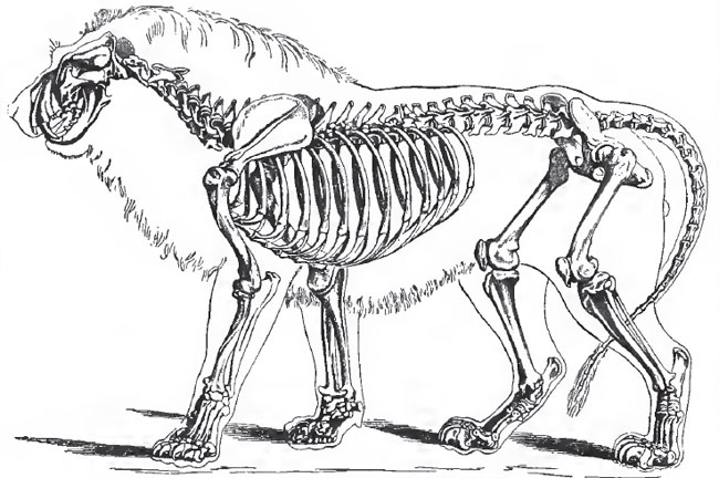 |
| Fig. 107. - The skeleton of Lion (Felis Leo) |
The olfactory lobes are usually large and the cerebral hemispheres elongated.
As the Dog (Canis familiaris) is an excellent and easily accessible example of a fissipede carnivore, it may be useful to mention some of the more important points in its anatomy.
The vertebral column contains twenty dorso-lumbar vertebra, of which thirteen are dorsal and seven lumbar, three sacral, and eighteen to twenty-two caudal vertrebrae. The atlas has broad and rounded alae, the anterior margins of which are deeply excavated near the roots. The posterior edge of the spinous process of the axis vertebra is almost perpendicular and very thick.
Nine pairs of ribs are usually connected by sterno-costal cartilages vdth the sternum, which is composed of eight laterally- compressed stenebrae. Only two of the three anchylosed sacral vertebrae articulate with the ilia.
As in the Carnivora in general, the occipital foramen is placed at the posterior end of the skull, and looks almost directly backward. The sagittal and lambdoidal crests are greatly developed and meet in a prominent occipital spine; the zygomata are very wide and arched outward; and the coronoid process of the mandible is very large. The size of these parts is in relation to the magnitude of the muscles of the neck and jaws.
The ramus of the mandible is nearly straight, the proper angle of the jaw being obsolete. A supra-angular process projects outward from the ascending portion of the ramus, and takes the place of the proper angle. The articular condyle is much elongated transversely, narrow and convex from before backward; and the pre-and post-glenoidal processes of the squamosal are produced downvrard so as to convert the joint into a complete ginglymus and to restrict the motion of the jaw to the vertical plane. The supra-orbital processes of the frontals are small and pointed. The root of the alisphenoid is traversed by a longitudinal canal. The tympanum is bounded below by a convex osseous wall, which is termed the bulla. It opens externally by the short external meatus, at the inner end of which is a circular elevation for the attachment of the tympanic membrane. A short distance internal to this frame for the membrane of the drum, a low crest rises from the floor of the bulla and imperfectly divides it into an outer and anterior portion which communicates with the Eustachian tube, and an inner blind spheroidal cavity which occupies the greater part of the bulla. The part of the bulla which forms the floor of this cavity is the result of the ossification of a process of the periotic cartilage, while the other part is furnished by the tympanic bone. The low crest is produced by the conjunction of both. Posteriorly and internally, the periotic region of the bulla presents a canal, through which the internal carotid artery passes. The posterior opening of the carotid canal looks into the foramen lacerurn posticum, and is not visible without dissection. There is a large paroccipital process, with a prominent free extremity; but, for the greater part of its length, it is closely applied to the back of the bulla. The condyloid foramen is quite distinct from the foramen lacerurn posterius. A large foramen behind the glenoidal cavity transmits a vein from the interior of the skull. In the nasal cavity, the ethmoidal turbinals are very large; the superior turbinals are prolonged into the great frontal sinus, and the inferior turbinals unite, in the middle line, with the septum.
The clavicles of the Dog are always rudimentary, and are generally represented only by a gristly intersection of the muscles which represent the sterno-mastoid and deltoid.
The olecranar fossa of the humerus is perforated. The hallux is much shorter than the other digits. When the Dog stands, the metacarpal bones of these digits are nearly vertical; the basal phalanges are horizontal; the middle and the distal phalanges are inclined in the form of a V with the apex (the articulation between the two) downward. The claws are, consequently, raised from the ground, the foot resting partly on a thick integumentary pad, which lies beneath the basal phalanges; and, partly, on the under surfaces of the joints between the middle and the distal phalanges. The distal phalanges are kept bent upon the middle ones by elastic ligaments, which pass from one to the other, and which antagonize the action of the long flexors. The Dog, therefore, possesses the mechanism for the retraction of the claws, but its action is not sufficient to protect them from wear. Fabellae or sesamoid bones developed in the tendons of the gastroenemius, lie behind the condyles of the femur. The fibula is thin and closely applied to, but not anchylosed with, the tibia. The hallux is usually rudimentary; only the metatarsal, and the basal phalanx, being represented by two small ossicles. In some breeds of dogs, however, the hallux is fully developed.
In the myology of the Dog the insertion of the tendon of the external oblique muscle of the abdomen presents some interesting peculiarities. The outer and posterior fibres of this muscle end in a fascia which is partly continued over the thigh as fascia lata, and partly forms an arch (Poupart's ligament) over the femoral vessels; by its inner end it is inserted into the outer side of a triangular fibro-cartilage, the broad base of which is attached to the anterior margin of the pubis, between its spine and the symphysis, while its apex lies in the abdominal parietes. The internal tendon of the external oblique unites with the tendon of the internal oblique to form the inner pillar of the abdominal ring, and is inserted into the inner side of the triangular fibro-cartilage. The pectineus is attached to the ventral face of the cartilage; the outer part of the tendon of the rectus into its dorsal face; but the chief part of that tendon is inserted into the pubis behind it. This fibro-cartilage appears to represent the marsupial bone, or cartilage, of the Monotremes and Marsupials.
The trapezius and the sternomastoid coalesce into a single muscle; and, in the absence of a complete clavicle, the outer fibres of the latter and those of the anterior part of the deltoid are continuous. In this way a muscle which has been called levator humeri proprius is formed. The omohyoid and the subclavius are absent. There is a trachelo-acromialis and a dorso-epitrochlearis. The supinator longus is absent, but there is a pronator quadratus. The extensor communis digitorum manus divides into four tendons, in which sesamoid bones are developed over the articulations between the first and second phalanges. The extensor primi internodii pollicis is absent. The extensor secundi internodii is one muscle with the extensor indicis. The extensor minimi digiti sends tendons to the third, fourth, and fifth digits. All these deep extensors have sesamoid bones over the metacarpo-phalangeal articulations. The palmaris longus appears to be absent; but all the other flexors of the manus, even the palmaris brevis, are represented. The tendons of the flexor pollicis longus aud flexor digitorum, perforans are united. The divisions which the common tendon sends to the five digits develop sesamoid bones, just before their insertions into the bases of the distal phalanges. The fifth digit has its abductor, flexor brevis, and opponens; the pollex, an abductor, adductor, flexor brevis, and, perhaps, an opponens. The second, third, and fourth digits have each a pair of flexores breves, which represent the interossei, and are inserted into the bases of the proximal phalanges, a relatively large sesamoid being developed in each. Each sends off a fine tendon dorsad to the extensor sheath. The plantaris is large, and, as in the Pig, its tendon passes into the representative of the flexor brevis digitorum pedis. The tendons of the flexor hallucis longus and flexor perforans unite into a common tendon, which subdivides into slips for the digits.
The dental formula of the Dog is i. 3.3/3.3 c. 1-1/1-1 p.m. 4.4/4.4 m. 2.2/3.3=42. The two upper inner incisors, on each side, have distinctly trilobed crowns-the lateral cusps of the crown arising from outgrowths of the cingulum at its base. The outer incisor is larger than the others, and its middle cusp is very large, while the outer is rudimentary. The large canine has a strong, curved, pointed crown, with a longitudinal ridge along its posterior face. The crowns of the anterior three premolars are triangular, with a smooth-cutting anterior edge; the hinder edge is also sharp, but is divided by a notch into two lobes, of which the hinder is the smaller. These teeth are two-fanged. The fourth premolar is a large tooth. In form, its crown has a general similarity to that of the foregoing; but, firstly, the posterior lobe is relatively much larger, and pointed, so as to form an obvious second cusp; and, secondly, a strong process of the crown projects inward from its anterior end, and is supported by a distinct fang-so that this premolar is three-fanged. It is termed a carnassial, or sectorial, tooth, as it bites like a scissors-blade against a corresponding tooth in the mandible. The preceding teeth have cutting crowns; but those of the molars are broad and crushing. They exhibit an outer division, formed by two large subequal cusps, and an inner division, also presenting two cusps, the posterior of which is much smaller than the anterior. In addition, the cingulum sends up a strong process on the inner side of the crown.
In the lower jaw, the crowns of the incisors, the outer of which is the largest, are all trilobed. The outer cusp is stronger than the inner in all, and particularly in the outer incisors. The canines resemble those of the upper jaw. Each premolar has two fangs and a sharp triangular crown, the posterior edge of which is trilobed, as in the upper premolars; but the posterior lobe is small in the fourth, which differs but little from the rest. The first molar, on the other hand, is a large tooth, with a blade-like crown, which bites against the inner side of the upper fourth premolar, and is called the carnassial or sectorial tooth of the lower jaw. The crown is elongated, and presents a large anterior external cusp, divided into two lobes by a deep notch. On the inner side of this is a small internal cusp. The two posterior cusps are very much lower than the anterior ones, and form a sort of heel to the blade-like anterior portion of the crown. An oblique ridge connects the outer and larger of the two posterior cusps with the small inner and anterior cusp. The second molar has a broad quadricuspidate crown, the inner posterior cusp being almost obsolete. The crown of the last molar is small, simple, and obtusely conical.
It thus appears that the sectorial, or carnassial, teeth in the two jaws differ in their nature, the upper being the last premolar, and the lower the anterior molar. The milk dentition of the Dog is d.i. 3.3/3.3 d.c. 1-1/1-1 d.m. 3.3/3.3, the "first premolar" of the adult dentition having no deciduous predecessor; so that, in this, as in so many other cases, it is doubtful whether it ought to be counted in the milk, or in the adult, dentition. The middle deciduous molar in both jaws resembles the hindermost premolar of the adult dentition, and the hindermost, the tirst molar of the adult. The so-called "first premolar" of the adult, and the anterior molars, appear before any of the deciduous molars are shed.
The caecum of the Dog is long, and folded upon itself, in which respects it is unlike that of other Carnivores. The arch of the aorta gives off an anonyma and a left subclavian.
In the brain, the olivary bodies are inconspicuous, the corpora trapezoidea large, and the corpora mammillaria distinctly double. The olfactory lobes are very large, and expand posteriorly on the sides of the brain into a broad mass continuous with the gyrus uncinatus, or hippocampal lobule. The cerebral hemispheres extend for a considerable distance over the cerebellum, in the upper view, and overlap it laterally. The Sylvian fissure does not extend more than half-way to the median fissure. The surface which answers to the insula is quite smooth. The anterior ends of the calloso-marginal sulci pass on the upper surfaces of the hemispheres, and give rise to the "crucial" sulcus. There are three principal gyri upon the outer surfaces of the hemispheres; one which immediately bounds the Sylvian fissure, one which runs along the upper margin of the hemisphere, and one between these two. The corpus callosum is long, and the anterior commissure well developed.
There is a musculus choanoides in addition to the usual ocular muscles, and the rudimentary nictitating membrane is said to possess a muscle.
The tensor tympani arises from a deep pit above the promontory, and its tendon passes directly outward to the malleus.
The male is devoid of Cowper's glands. The penis has a bone, and the glands becomes swollen during copulation, so as to prevent the withdrawal of the penis from the vagina of the female. The ovary of the female is enclosed in a sac of the peritonaeum, and the uterus has long cornua. The umbilical sac is drawn out to a point at each end.
The Dogs (including the Wolves, Jackals, and Foxes, under this head) form the most central group of the Carnivora, which may be termed the Cynoidea (See Prof. Flower's important memoir on tlie Classifleation of the Carnivora in the Proceedings of the Zoological Society for 1869.) From these the Bears, Weasels, and Procyonidae depart, on the one hand, and the Cats, Civets, and Hyeenas on the other. The former group (Arctoidea) have the cavity of the bulla tympani undivided by a septum. The paroccipital process is not applied to the posterior wall of the bulla. The mastoid process is widely separated from the paroccipital. The condyloid foramen is not merged in a common opening with the foramen lacerum posticum. The intestinal canal is devoid of a caecum. The large penis has a bone which is not grooved; there are no Cowper's glands, and the prostate is small.
In the latter group (Ailuroidea) the bulla tympani is large and rounded, and the septum, which is rudimentary in the Cynoidea, is so much enlarged as to leave only a narrow aperture of communication between the two chambers. The par-occipital is closely applied to the posterior wall of the bulla. The mastoid process is often obsolete. The condyloid foramen opens into a fossa common to it and the foramen lacerum posticum. All have a short caecum. The penis is small, and its bone small, irregular, or absent. They have Cowper's glands and a well-developed prostate.
The Cynoidea are all digitigrade, and resemble the Dog in their dentition. The Arctoidea are plantigrade, while the Ailuroidea are for the most part digitigrade, but may be plantigrade. In dentition, each of these groups presents forms such as the Bears on the one hand, and the Cats on the other, which may be regarded as extreme modifications, in opposite directions, of the type exhibited by the Dog.
In the Bears, the dental formula is the same as in the Dogs, but the crowns of the teeth are all more obtuse. The sectorial teeth lose their marked characters, and the molars have flat and tuberculated crowns. The anterior premolars fall out as age advances. It is a remarkable circumstance that the teeth of frugivorous and carnivorous Bears exhibit no such differences as would lead to a suspicion of their complete difference of habit, if we were acquainted with these animals only in the condition of fossils.
The Cats have the dental formula i. 3.3/3.3 c. 1-1/1-1 p.m. 3.3/2.2 m. 1.1/1.1=30. The canines are very long and sharp. The premolars are like the Dogs', except that they are sharper, and that the hindermost (the sectorial tooth) has hardly any internal process. The single upper molar is a small tooth with a flat, transversely-elongated crown, and it lies within, as well as behind, the great sectorial premolar. In the lower jaw, the sectorial, or first, molar is the last tooth in the series. The crown is a deeply-bifnrcated blade representing the anteroexternal cusp of the corresponding tooth in the Dog. The "heel" is obsolete.
While the Bears are among the most completely plantigrade of the Carnivora, the Cats are most entirely digitigrade, and the apparatus for the retraction of the ungual phalanges is so well developed that the claws are completely retracted within sheaths of the integument, when the animal does not desire to use them. To this end the elastic ligaments are very strong, and the median phalanx is excavated, in order to allow of the lodgment of the retracted phalanx on one side of it.
b. The Pinnipedia, or Seals and Walruses, are those Carnivora which come nearest the Cetacea. The tail is united, by a fold of skin which extends beyond its middle, with the integument covering the hind-legs. These are, in most species, permanently stretched out in a line with the axis of the trunk. The pinna of the ear is small or absent. The toes are completely united by strong webs, and the straight naila are sometimes reduced in number, or even altogether abortive. The inner and the outer digits of the pes are very large. The incisors vary in number and lose their cutting form. The pre molar and molar teeth are similar in character, and never have more than two fangs. There is no lachrymal bone or canal.
The brain-case of the cranium is generally much more rounded than that of other Carnivora; and, in some genera, the supra-orbital processes of the frontals are very largely developed. In both of these characters, and in the great breadth and complication of the convolutions of their cerebral hemispheres, as well as in their relatively small olfactory nerves and anterior commissure, the Pinnipedia approach the Cetacea.
There are three groups of Pinnipedia: the Otaridae, the Trichechidae, and the Phocidae.
1. The Otaridae, or Eared Seals, are so termed because the ear possesses a distinct though almost rudimentary pinna. These Seals have long necks, and can stand or walk upon all fours, the hind-limbs being capable of supporting the body in the ordinary way.
In many respects, these animals are closely allied with the Bears; and by no part of their organization is this more clearly shown than by the skull, which in its general form, its large supra-orbital processes, the small and rugged bulla tympani, the perforation of the alisphenoid by a canal, and the presence of a crest on the inner surface of the parietals, is extremely ursine.
2. The Trichechidae, or Walruses, are devoid of external ears, but resemble the Otaridae in their mode of standing and walking. The skull resembles that of the Bear in the same respects, but the muzzle is distorted by the enormous development of the superior canines. The Walruses resemble the Bears in another point, namely, in the presence of a supplementary bronchus; the right bronchus, before it reaches the lung, dividing into two trunks, a large and a small. The thyroid cartilage is deeply excavated, in front, by a triangular fissure; and the epiglottis is extremely small.
In the brain, the remarkably large and richly convoluted hemispheres cover the cerebellum, and present a rudimentary posterior cornu. The anterior commissure is very small, as are the olfactory nerves.
The dentition of the Walrus is extremely peculiar. In the adult, there is one simple conical tooth in the outer part of the premaxilla, followed by a huge tusk-like canine, and three, short, simple-fanged teeth. Sometimes, two other teeth, which soon fall out, lie behind these, on each side of the upper jaw. In the mandible there are no incisors, but a single short canine is followed by three, similar, simple teeth, and by one other, which is caducous.
The dental formula is therefore i. 1-1/0.0 c. 1-1/1.1 p.m.m. 3-3/3.3 + 2.2/1.1=24.
3. The Phocidae, or ordinary Seals. - The pinna is altogether absent. The hind-limbs are permanently stretched out, parallel with the tail; and, consequently, they are unable to support the body, or assist in locomotion on land.
The space between the orbits is extremely narrow, and supra-orbital processes are absent. The bulla tympani is very large and thick-walled; and the middle are much shorter than the outer digits of the pes.
The common Seal (Phoca vitulina) is a native and accessible member of this group. It has a rounded head and a neck which is well mai-ked, though shorter in proportion than that of the Eared-seals. The nasal apertures are slit-like and can be closed at will, the eyes large and brilliant, and the auditory apertures small and devoid of a pinna. The limbs are large, and their distal longer than their proximal divisions. The fore-limb is buried beyond the elbow in the common integument, but the flexible wrist allows the weight of the body to be supported by the palmar surface of the manus. The hind-limbs, on the contrary, are permanently extended and turned backward parallel with the tail, which lies between them, and with which they form a sort of terminal fin. When the Seal swims, in fact, the fore-limbs are applied against the sides of the thorax, and, the hinder moiety of the body being very flexible, the conjoined hind-limbs and tail are put to the same use as the caudal fin of a Cetacean. The Seal has twenty dorso-lumbar vertebrae, of which five are lumbar. There are four sacral vertebrae, but only one of these unites with the ilia. Eleven vertebrae enter into the formation of the short tail. There are ten true ribs and nine sternebrae, the manubrium being prolonged forward into a long cartilaginous process.
The brain-case is smooth, rounded, and spacious, but the cranium narrows rapidly in the interorbital region. Its floor is remarkably flattened from above downward and very thin, the broad basi-occipital sometimes presenting a perforation in the dry skull. The falx is partially, and the tentorium is wholly, ossified. The occipital segment is very large, and the supraoccipital advances between the parietals, but does not separate them completely. The alisphenoids are small and almost horizontal, and the synchondrosis between the basisphenoid and presphenoid persists. In all these respects the Seal's skull is strikingly cetacean. In fact, if the supra-orbital processes were sawn off, a Porpoise's brain-case would closely resemble a Seal's. But the nasal bones and the parietals are large, and the ethmoidal region is very peculiar. The lamina perpendicularis is largely ossified, and the vomer soon becomes ossified into one mass with it. The two ethmoidal turbinals (or the superior and middle) are small and flattened, and the latter anchyloses with the vomer on each side. The inferior, or maxillary, turbinal is extremely large and complicated, and it blocks the nasal passage in front of the others like a sieve, or strainer. There is no lachrymal bone, but the jugal is large. The squamosal is anchylosed with the periotic and tympanic. The latter is massive and shell-shaped, somewhat as in the Cetacea, but it has rather diiferent relations to the auditory meatus. The periotic is very large, and its tumid pars mastoidea appears largely on the exterior of the skull. The fossa under the superior vertical semicircular canal is prolonged into this tumid part of the periotic.
The alveolar portions of the premaxillae are very small, but these bones extend far up the sides of the anterior nares. The maxillae do not extend over the frontals. The mandible has a well-developed coronoid process.
The pollex is the longest and strongest digit, the others gradually decreasing in length. The fifth metacarpal articulates with the cuneiform bone, as well as with the unciform.
The ilium is short, and the long pubis and ischium are greatly inclined backward, so that the long diameter of the os innominatum makes only an acute angle with the spine. The femur is much shorter than the humerus. The tibia and fibula are anchylosed, and more than twice as long as the femur. The pes is longer than the tibia. The astragalus has a peculiar, roof-shaped, tibial surface, and sends a process backward which contributes to the formation of the very short heel. The hallux is the strongest of the digits; while this and the fifth digit are the longest of those of the pes.
The cutaneous muscle is largely developed and inserted into the humerus. The pectoralis major is very large, and arises from each side of the prolonged manubrium, and even in front of it, beneath the neck; the fibres of the muscles of opposite sides are continuous. The palmaris longus is a strong muscle, but the proper digital muscles are weak or absent, as in the case of the abductor, adductor, flexor brevis, and opponens of the fifth digit. A special long abductor of this digit, however, passes from the olecranon to the distal phalanx. The iliacus is wanting, and there is no psoas major; but muscles which represent the psoas minor and the subvertebral muscles of the Cetacea are very large and play an important part iu effecting the locomotion of the Seal. The pectineus is very small, and the other adductors are inserted, not into the femvir, but into the tibia. The glutaeus maximus is inserted into the whole length of the femur. The semi-membranosus and semitendinosus are replaced by a caudo-tibialis, which arises from the anterior caudal vertebrae and is inserted into the tibia, some of its tendinous fibres extending to the plantar aspect of the hallux. The poplitaeus and gastrocnemius are strong, but there is no solaeus. The tendon of the plantaris passes over tiie calcaneum and ends on the plantar fascia of the perforated tendon of the fourth digit. The other perforated tendons seem to arise from the fascia attached to the calcaneum.
The dental formula is i. 3-3/2-2 c. 1-1/1-1 m.p.m. 5-6/5-6=34.
The grinding teeth have triangular crowns with notched edges, and at most two fangs.
The milk-teeth are shed during foetal life, and at this period there are three molars above and below on each side, which appear to be replaced by the second, third, and fourth of the adult set. If such be the case, only the hindermost of these last will be a true molar.
The tongue is bifid at the extremity. The oesophagus, very wide and dilatable, passes without any very well-marked line of demarcation into the stomach, which is a great pyriform sac with its pyloric end bent upon itself. The intestine is about twelve times as long as the body. The colon is short, and is provided with a caecum. The liver is divided into a great number of lobules, which are, as it were, set upon the inferior cava. The latter vessel, just below, the diaphragm, presents a great dilatation, into which the venae hepaticae of the several lobules open. After traversing the diaphragm, the vena cava is surrounded, for about an inch, by a layer of red circular muscular fibres. The aorta and the pulmonary artery are both dilated at their commencements.
The penis of the male is contained within a prepuce, suported by a loop of the cutaneous muscle. There is a large os penis, which presents a groove for the urethra inferiorly. The prostate is small, and there are no vesiculae nor Cowper's glands. The testes lie just outside the inguinal canal. The anus and the vulva of the female are surrounded by a common fold of integument. The clitoris has no bone. The body of the uterus is divided by a longitudinal septum.
Support our developers


