The Chelonia
The Tortoises and Turtles are those reptiles which most nearly approach the Amphibia, though they depart very widely not merely from the amphibian, but from the ordinary vertebrate type, in some respects.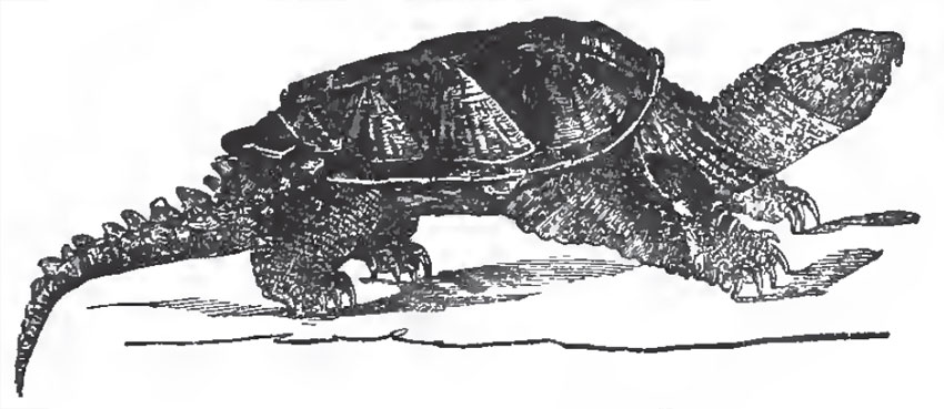 |
| Fig. 61. - The Alligator Terrapene (Chelydra Serpentuna). |
The dermal ossifications may best be described in connection with the endoskeleton.
The presacral vertebrae are few in number. In the Green Turtle (Chelone midas) there are eight cervical, and ten dorsal, in front of the sacrum, which is composed of two vertebrae. In all the cervical vertebrae the neurocentral sutures persist; there are no transverse processes, or ribs, and the spines are low or obsolete. The first vertebra, or atlas, is a ringlike bone, composed of three pieces, one basal and two superolateral. The second is a true axis vertebra, the central part of the centrum of the atlas ossifying apart, as an odontoid bone, and attaching itself to the front face of the centrum of the second vertebra.
The other cervical vertebrae are remarkable for the singular variety in the disposition of their articular cups and balls.
Thus the third is opisthocoelous; the fourth, biconvex; the fifth, procoelous; the sixth, also procoelous, but the posterior face is nearly flat, and very broad; in the seventh, both the anterior and the posterior faces are very broad and flattened, the posterior being the more convex. The eighth cervical vertebra is procoelous, and differs from the rest by the expansion of its neural spine, and by the arching backward of its postzygapophyses over the convex prezygapophyses of the first dorsal vertebra, upon which the former play backward and forward.
All the cervical vertebrae are very freely movable upon one another, and confer great flexibility on the neck. In striking contrast with this arrangement, the ten following vertebrae have flattened faces, firmly united by cartilage. If any one of these vertebrae, from the second to the ninth, be examined, it will be found that the elongated centrum is only loosely united with the neural arch, and that the summit of the neural arch is continuous with a broad flat plate of bone, which forms one of the eight median elements of the carapace, or neural plates (Fig. 62, V).
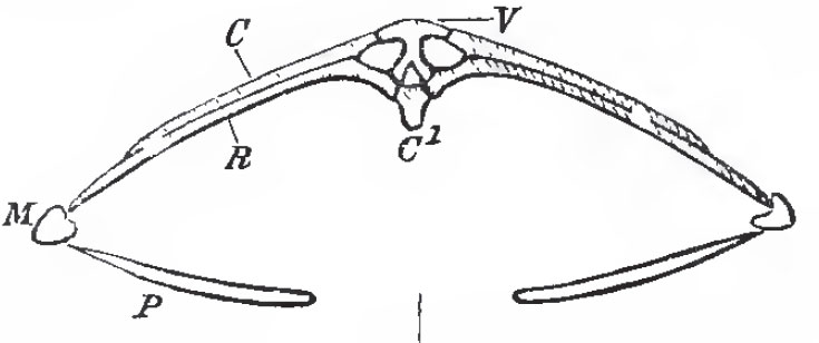 |
| Fig. 62. - Transverse section of the skeleton of Chelone midas in the dorsal reg:ion: C1, centrum: V, expanded neural plate; C, costal plate; R, rib; M, marginal plate; P, lateral element of the plastron |
The first dorsal vertebra differs from the others in many respects. The anterior face of its centrum is concave, and looks downward and forward, while its prezygapophyses are much prolonged, in order to articulate with the convex posterior face of the centrum and prolonged postzygapophyses of the last cervical vertebra. The spinous process of this vertebra does not pass into the bony nuchal plate of the carapace, which lies above it (Fig. 63, Nu), and its rib does not expand into a costal plate, but becomes connected with the costal plate of the second dorsal vertebra. The neural arch of this vertebra is shorter, from before backward, than its centrum; and the neural arch of the second dorsal vertebra extends forward and overlaps the centrum of the first, for the space thus left unoccupied. The rib of the second vertebra is also carried forward, and articulates not only with its own centrum and neural arch, but with the posterior edge of the centrum of the first vertebra.
 |
| Fig
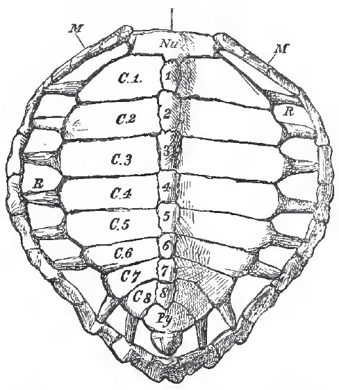
63. - Dorsal view of the carapace of Chelone midas: Nu, nuchal plate; M, marginal
plates; R, ribs; 1-8, neural plates; C, 1-C.8, costal plates; Py, pygal plates. |
These arrangements are repeated by the other dorsal vertebrae and ribs, up to the ninth inclusive; but, in the tenth, the neural arch occupies only the anterior half of the centrum of its own vertebra, and the rib is very small, and has no costal plate.
The union of the neural and costal plates of the eight dorsal vertebrae, from the second to the ninth inclusively, gives rise to the principal part of the carapace, or dorsal moiety of the bony shell of the Chelonian. The first and the tenth dorsal vertebrae contribute nothing to the carapace, their small ribs merely becoming attached to the costal plates behind and before them.
In front of the first neural plate, and joined with it by a serrated suture, lies a large nuchal plate (Fig. 63, Nu), which forms the anterior median boundary of the carapace. This nuchal plate sends down from its under-surface a median process, which is joined by ligament with the expanded neural spine of the eighth cervical vertebra. Behind the eighth neural plate, three other median pygal plates (Fig. 63, Py) succeed one another. The anterior two of these are united by sutures with the eighth neural and costals, and with one another; but the third is connected externally only with the marginal plates. All three are perfectly distinct from the subjacent vertebrae.
The sides of the carapace are completed, between the nuchal and pygal plates, by eleven marginal plates (Fig. 63, M) on each side. Eight of these receive the ends of the ribs of the second to the ninth dorsal vertebrae, in the way already described.
There is no doubt that the nuchal, the pygal, and the marginal plates of the carapace are membrane-bones, developed in the integument, quite independently of either the vertebrae or the ribs. But it appears that the neural plates and the costal plates exist, as expansions of the cartilages of the Neural spines and ribs of the primitive vertebrae, before ossification takes place. This being the case, the neural and costal plates are vertebral and not dermal elements, however similar they may seem to be to the nuchal, pygal, and marginal plates. But this ultimate similarity of bones of totally distinct origin is not more remarkable here than in the case of the skull, where the parietal and frontal bones stand in the same relation to the supra-occipital bone as the nuchal and pygal plates do to the neural plates of the carapace.
 |
| Fig. 64. - The plastron of the Green Turtle (Chelone midas): I.cl, interclavicle; cl, clavicles; Hy.p., hyoplastron; Hp.p., hypoplastron; Xp., xiphiplastron. |
There are no sternal ribs, and no trace of a true sternum has yet been discovered in the Chelonia. The plastron is wholly composed of membrane-bones, which are developed in the integument, and lie, in part, in front of, and, in part, behind, the umbilicus of the foetus. The latter, at least, therefore belong to the abdomen, and the plastron is a thoracicoabdominal structure.
In the turtle the plastron consists of nine pieces-one median and anterior, four lateral and paired (Fig. 64). These pieces may be named-the median, entoplastron; the first lateral, epiplastron; the second, hyoplastron; the third, hypoplastron; and the fourth, xiphiplastron.(Believing the plastron to answer to the sternum of other Vertebrata anatomists have termed these elements of the plastron entosternum, episternum, hyosternum, hyposternum and xiphisternum.) The entoplastron and the two epiplastra correspond with the median and lateral thoracic plates of the Labyrinthodont Amphibia, and very probably answer to the interclavicle and clavicles of other Vertebrata.
The sacrum consists of two vertebrae. The expanded sacral ribs are not anchylosed with the centra and arches of their vertebrae.
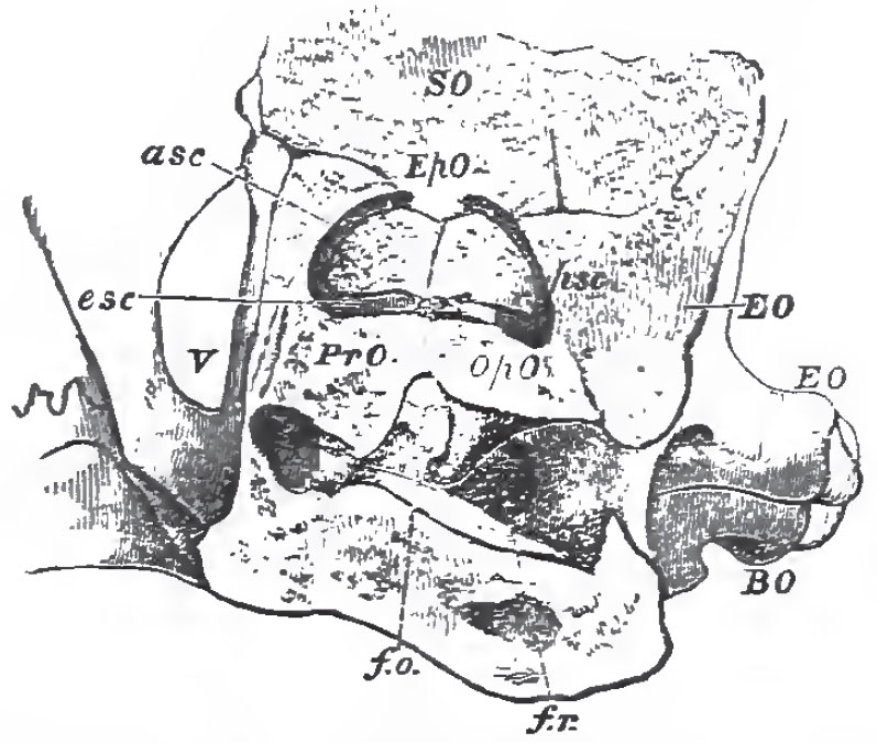 |
| Fig. 65. - External view of a section of the anditory region of the skull in a Turtle (Chelone midas): f. o., fenestra ovalis: f. r., fenestra rotunda; esc, asc, psc, external, anterior, and posterior semicircular canals. |
In the skulls of the Chelonia all the bones, except the mandible and the hyoidean arch, are immovably united together.
In the occipital segment of the adult, the supra-occipital is united with the epiotic, but the ex-occipital usually remains perfectly distinct from the opisthotic. The basisphenoid is large and distinct. The alisphenoidal region remains unossified; but the large parietals send down a prolongation on each side, which plays the part of an alisphenoid. Neither the presphenoid nor the orbitosphenoids are represented by bone, but there are large frontals. In the periotic capsule the large prootic and the opisthotic (Cuvier's occipitale externe) remain distinct bones, but the epiotic unites with the supraoccipital.
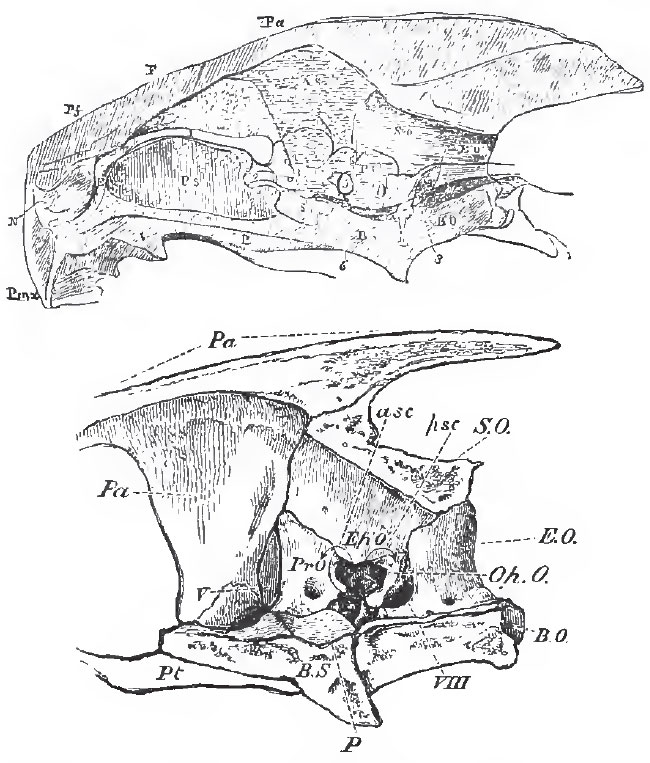 |
| Fig. 66. - Longitudinal sections of the skull of the Turtle. The upper figure represents the entire skull with the outline of the brain in situ; the lower gives a larger view of the inner face of the bones of the posterior moiety of the skull. |
The naso-ethmoidal cartilage largely persists; but it becomes covered above and at the sides by a large bone, which meets with its fellow in the middle line, and occupies the position of the lachrymal, prefrontal, and nasal. The premaxilla are small, and usually united together. There is a single vomer, produced downward into a median internasal plate, which expands below, and joins the palatine plate of the palatine bone.
Above the posterior and upper part of the orbit lies a postfrontal, and, behind this, a squamosal is placed at the sides of the periotic capsule, and above the large quadrate bone. The postfrontal and spuamosal occupy the upper part of the temporal region of the skull. Below these, a quadratojugal and a jugal connect the quadrate bone with the large maxilla.
In some genera, as Chelone and Chelydra, the skull possesses a sort of false roof, formed by the expansion of a median ridge, developed from the parietal bones, into a broad plate, which becomes suturally united with the postfrontals and squamosals.
The quadrate bone is firmly fixed to the sides of the periotic region of the skull, and ends below in a strong condyle for the mandibles. The long and broad pterygoid bones unite with one another in the middle line, and are firmly fixed to the base of the skull, as in Plesiosauria and Crocodilia. They unite only with the upper part of the quadrate bone, as in the latter reptiles.
The palatines are firmly united with the pterygoids, behind, and with the vomer above and in front. They are prolonged downward, and develop a short palatine plate, which unites with the produced and expanded lower edge of the vomer, to bound the posterior nares. (Fig. 67, Vo, N1.)
The dentary pieces of the two rami of the mandible are represented by one bone, as in Birds.
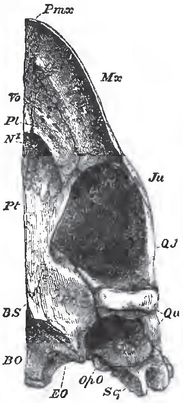 |
| Fig. 67. - The left half of the underside of the skull of a Turtle; N1, posterior nares. |
The pectoral and pelvic arches appear, at first sight, to have a very anomalous position in the Chelonia, inasmuch as they seem to be situated inside, and not outside, the skeleton of the trunk. But since the plastron does not answer to the sternum of other Vertebrata, but to part of the dermal skeleton, the anomaly does not really exist on the ventral side. And, as to the dorsal side, the pectoral and pelvic arches of the foetal Chelonian are at first situated in front of, or behind, and external to, the ribs, as in other Vertebrata. It is only as development advances, that the first costal plate extends over the scapula, and the hinder costal plates over the ilium.
The pectoral arch is ossified in such a manner that the scapula and precoracoid form one bone, while the coracoid remains distinct. The free ends of the coracoid and precoracoid are usually connected together by a fibro-cartilaginous band, representing the epicoracoidal cartilage in Lacertilia. There is no clavicle, unless the epiplastra and entoplastron represent that bone.
The carpus of the Chelonia contains nine primary ossicles, as in the Urodela-three in the proximal row, one central, and five distal-and these almost always remain distinct.
There are five digits, the numbers of the phalanges of which present no constancy.
The pelvis contains the usual bones. The pubes (which are very large) and the ischia meet respectively in a long symphysis; and, sometimes, the foramina obturatoria are completed, internally, by the meeting of the bony pubes and ischium of each side in the middle line.
The pelvis is not usually united with either the carapace or the plastron, but in Chelys, Chelodina, and some other genera, the ilia unite by synchondrosis, or anchylosis, with the last costal plate, and the pubis and ischium with the xiphisternal plates, so that the pelvis becomes firmly fixed between the carapace and plastron.
The proximal row of the tarsal bones consists usually of an astragalus, formed by the union of the tibiale and intermedium, and of a fibulare or calcaneum. In Chelydra there is a centrale. In Chelone, Emys, Testudo, and Trionyx the centrale is united with the astragalus; and in Emys, the calcaneum coalesces into the astragalus, so that the proximal portion of the tarsus consists of one bone. In the distal series the two fibular tarsals are united into a cuboid bone. There are five digits, and the fifth metatarsal has a peculiar form, as if bent upon itself at right angles, in the middle of its length.
In the Testudinea there are only two phalanges in each digit of the pes.
The Chelonia are divisible into the Testudinea, the Emydea, the Trionychoidea, and the Euereta.
1. The Testudinea have the horny jaws naked and cutting, or denticulated. The eyes are lateral, the tympanic membrane is exposed, the short and thick limbs have the toes (all of which have nails) bound together by the integument. The horny plates of the carapace and plastron are well developed.
The Land Tortoises belong to this division. The carapace is usually very convex, and sometimes (as in the genus Pyxis) the anterior part of the plastron is movable, and can be shut up like a lid. In Cinyxis, the hinder part of the carapace is similarly mobile.
2. The Emydea have, usually, horny cutting jaws, uncovered by lips; the tympanum exposed, and the limbs more slender than in the Testudinea, with five-clawed digits, which are only united by a web. The homy plates of the carapace and plastron are well developed.
These are the River and Marsh Tortoises. They are further divisible into two groups, in the one of which, the Terrapenes, the pelvis is free, the neck bends in a vertical plane, and the head, is almost completely hidden by the carapace when retracted (Emys, Cistudo, Chelydra). In Cistudo, Cinostenum, and Staurotypus, the hinder part of the plastron is mobile. In the other division, the Chelodines, the pelvis is fixed to the carapace and plastron, the neck bends sideways, and the head cannot be completely retracted under the carapace (Chelys, Chelodina.)
3. In the Trionychoidea (Mud or Soft Tortoises), the jaws have an external cutaneous lip; the nasal organ is prolonged into a kind of snout, and the head is covered by a soft skin without any visible tympanic membrane. The limbs are flattened, somewhat finlike, and pentadactyle; but only three digits have nails. The integument develops no horny plates, but is quite soft. Tlie costal plates are shorter than in other Chelonia, and the marginal ossicles are either rudimentary or absent.
The genera Gymnopus, Cryptopus, and Cycloderma, constitute this division; they all inhabit the fresh waters of hot latitudes.
The Euereta, or Turtles, have an exposed, hooked, horny beak, with a blunt snout. The tympanum is hidden by the integument. The limbs, of which the anterior pair are much the longer, are converted into paddles, the digits being much flattened and elongated, and immovably united together by the integument; only one or two nails are developed. The skin of the body is either rugose (Sphargis), or covered with thick epidermic plates (Chelone.)
The two genera composing this group inhabit the seas of warm climates.
The Chelonia are first known to occur, with certainty, in the Lias. The older forms are, in many respects, intermediate between the Euereta and the Trionychoidea, but present no approximation to any other order of Reptilia.
Support our developers


