Growing Madin-Darby Canine Kidney Cells for Studying Epithelial Cell Biology
Epithelial cells display a structural and functional polar organization (Simons and Fuller, 1985). In these cells, the plasma membrane can be divided into two distinct domains, the apical membrane and the basolateral membrane, each containing different sets of proteins. The apical membrane facing a secretory or an absorptive lumen is delimited by a junctional complex from the basolateral membrane. The tight junction (zonula occludens) is the most apical member of the complex. It is found at the intersection between the apical and the lateral plasma membranes and joins each cell to its neighbors, thus limiting the diffusion of molecules between the luminal and the serosal compartments (Gumbiner, 1987). This junction also prevents the lateral diffusion of membrane proteins from one domain to another, thus maintaining their unique composition. Immediately basal to the tight junctions is the intermediate junction (zonula adherens or belt desmosomes). The other more basal junctional elements are desmosomes (maculae adherentes) and gap junctions, which attach the lateral membranes of adjacent cells to each other. The junctional complex is involved in sealing the epithelium; it prevents molecules from diffusing between adjacent cells. The basolateral membrane faces the bloodstream and is involved in cellcell contact and cell adhesion to the basement membrane.
For most studies on epithelial cell polarity, cultured cells have been used. These cells are superior to cells obtained from tissues because they can be grown under carefully controlled conditions and are easily manipulated. The cell population is homogeneous. Biosynthetic experiments using pulse-chase techniques with radioactive precursors can be accomplished at an analytical level with a short time resolution. Endocytosis and transcytosis can also be studied.
These problems can be overcome simply by growing the epithelial cells on permeable supports, such as polycarbonate and nitrocellulose filters. Epithelial cells form monolayers with a higher degree of differentiation when the basolateral surface is directly accessible to the growth medium. This is evident from the morphology of the cells, their increased responsiveness to hormones, and the exclusion of basolateral proteins from their apical surfaces.
Minimal essential medium with Earle's salt (MEM) is purchased as a powder (Cat. No. 11700-077) from Biochrom, mixed with Milli-Q-filtered H20, and sterile filtered. Glutamine (200mM, Cat. No. 25030-024), penicillin (10,000 IU / ml)-streptomycin (10,000 mg/ml) (Cat. No. 15140-122), trypsin (0.05%)-EDTA (0.02%) (Cat. No. 25300-054), and phosphate-buffered saline (PBS, Cat. No. 041-04040H) are from GIBCO-BRL. The Transwell polycarbonate filters (2.45 cm, Cat. No. 3412, and 10cm, Cat. No. 3419) are from Costar. Tissue culture flasks (75cm2, Cat. No. 156499) are from Nunc. The glass petri dishes (140mm diameter and 30mm high) for holding six 2.4-cm Transwell filters are from Schott Glasware. The laminar flow hood (Steril-Gard Hood Model VMB-600) is from Baker. The CO2 incubator (Model 3330) is from Forma Scientific. The inverted Diavert microscope is from Leitz. The electrical resistance measuring device (EVOM) is from World Precision Instruments. The centrifuge (Type 440) is from Hereaus-Christ.
III. PROCEDURES
A. Growing Madin-Darby Canine Kidney Cells on Plastic
MDCK I and II cells are passaged every 3-4 days up to 25 passages. One flask is usually split into five new flasks. MDCK II cells usually form domes within 2 days of splitting, whereas MDCK I cells do not blister.
Solutions
- MEM growth medium:
- Phosphate-buffered saline
- 0.05% trypsin/O.02% EDTA
| Stock | Volume/liter | |
| 5% fetal calf serum (MDCK II) | 100% | 50ml |
| 10% fetal calf serum (MDCK I) | 100% | 100ml |
| 2 mM glutamine | 200 mM | 10 ml |
| 100 IU / ml penicillin | 100X | 10ml |
| 100 / µg/ml streptomycin |
Steps
- Wash hands and wipe laminar flow hood with 70% ethanol. Warm all solutions to 37°C. All manipulations are done in the laminar flow hood. When splitting the cells, remove the growth medium from the 75-cm2 flasks containing the confluent layer of MDCK cells and add 10ml PBS. Rinse and discard wash solution.
- Add 5 ml trypsin-EDTA solution, seal flask, and incubate for 10-15min at room temperature (until small patches of cells are rounded up but not yet detached from the flask).
- Remove the trypsin-EDTA solution and add 1.5ml of fresh trypsin-EDTA. Reseal the flask and incubate at 37°C for 10-15 min (MDCK II) or 25-30 min (MDCK I). At this point the cells should flow down to the bottom of the flask when the flask is turned up. Hit the flask hard against the palm of your hand.
- Add 10ml prewarmed MEM growth medium and resuspend the cells with a sterile 10-ml pipette (at least five times up and down). Using the inverted microscope, check that the cells are not sticking to each other.
- Plate 2 ml of the cell suspension in a new 75-cm2 flask containing 20 ml of MEM growth medium.
B. Seeding MDCK Cells on Polycarbonate Filters
Solutions
- MEM growth medium containing 10% fetal calf serum, penicillin-streptomycin, and 2 mM glutamine (see solution A1).
- Phosphate-buffered saline
- 0.05% trypsin/O.02% EDTA
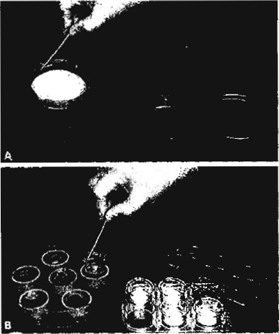 |
| FIGURE 1 (A) The glass petri dish contains one filter holder for one Transwell 2419 filter. (B) The glass petri dish contains six filter holders for Transwell 3412 filters. |
- Seed the cells on the filters at high density, higher than that achieved by confluent cells on plastic. The cells form tight junctions within 24h and reach maximum tightness on the filters in 4 days. During this time cell density increases to more than five times that achieved on plastic. We place the filters in the petri dishes containing growth medium. For seeding we use one 75-ml flask, containing a confluent layer of MDCK I and II cells, for 2.4-cm-diameter filters (Transwell 3412). If you use large 10-cm-diameter filters (Transwell 3419), seed one 75-cm2 culture flask of MDCK cells into each large filter.
- Pour off medium from the culture flask and rinse cells with 10 ml of warm PBS. Pour off PBS.
- Add 5 ml of warm trypsin-EDTA to cells. Leave in laminar flow hood.
- After 15min remove the trypsin-EDTA with a pipette, add 1.5 ml of trypsin-EDTA, and put the flask into a CO2 incubator (37°C) for 10-15min (MDCK II) or 25-30 min (MDCK I).
- Remove flask (cells should be loose). Hit the flask hard against your palm. Add 10ml of warm growth medium and suspend cells by pipetting up and down with a 10-ml pipette. Put suspension into a 50-ml Falcon tube and centrifuge for 5 min at 1000rpm in a Hereaus-Christ centrifuge.
- Remove supernatant and suspend cells in 9.5 ml of growth medium.
- Pour 90 ml medium into glass petri dish containing six Transwell 3412 filters. Use 140ml for one Transwell 3419 filter. The petri dishes contain filter holders specially made to fit either 3412 or 3419 filters (Fig. 1). Autoclave these units before use. Place filters into filter holders and allow filters to get wet from the bottom with medium. This should be done while the cells are in the centrifuge.
- Add 1.5 ml of cell suspension to each filter in its holder. Use six Transwell 3412 filters or one Transwell 3419 filter per petri dish. Be careful not to spill cells over the edge of the filter holder.
- Swirl petri dish gently to remove any trapped air from beneath filters.
- Place the petri dish with the filters in the CO2 incubator.
- Leave for 3-4 days in the incubator. No medium change is required during this time.
Steps
- Transepithelial resistance of filter-grown MDCK cells is measured with EVOM "chop-stick" electrodes. Each leaf has an outer and an inner electrode. The outside electrodes are small silver pads for passing current through the membrane sample. Inside the electrodes are small Ag/AgC1 voltage sensors.
- To test the instrument, switch the mode switch to R and turn the power on. Push the test R button. With the range switch in the 2000-V position, the meter will read 1000 (±1 digit). In the 20-k range, the meter will read 1.00. The meter is now ready for use.
- To test the electrodes, insert the small telephonetype plug at the end of the chopsticks electrode cable into the jack on the front panel of the EVOM. Place the tips of the electrodes into 0.1M KCl. Switch the mode switch to Volts. Turn the power switch on. The digital panel meter may read I or 2 mV due to the asymmetry of the voltage sensor pair. After 15min, adjust this voltage to 0mV with the screwdriver adjustment labeled "Zero V."
- Measure resistance. The electrode set is designed to facilitate measurements of membrane voltage and resistance of cultured epithelia in culture cups by dipping one stick electrode inside the cup on top of the cell layer and the second stick electrode in the external bathing solution. To measure resistance, immerse the electrode pair again into the electrolyte and set the mode switch to "Ohm." The display should read zero; if not, adjust the display to zero with the Ohms Zero screwdriver adjustment. Push the measure R button. A steady ohm reading of the resistance should result.
- When moving the electrodes from one dish to another it is best not to rinse the electrodes with distilled water. If it is necessary to wash the electrodes between measurements, they should be rinsed with the membrane perfusate (e.g., PBS). Do not touch the cell layer with the internal electrode when making a measurement. Small differences in the apparent fluid resistance may occur if the depth to which the electrodes' tips are immersed varies. If the tips are unusually dirty, a light and very brief sanding with a fine nonmetallic abrasive paper will clean the sensor tip. For sterilization the electrodes may be soaked in alcohol or bactericides. After sterilization, the electrodes should be rinsed extensively with sterile perfusing solution or 1 M KCl.
| Example | |
| a. Measure resistance R from solution + sample membrane support | 109 |
| b. Measure resistance R from solution + membrane support + tissue | 189 |
| c. Subtract (a) from (b) | 189- 109- 80, R (tissue) - 80 V |
| d. Calculate resistance × area product | resistance × area = 1.2 cm × p × r2 = 80V × 3.14 × (1.2cm)2 = 361.9 V cm2 |
The cell layer on the filter cannot be observed in the inverted microscope because the filters are not transparent. Transparent filters are also available commercially' but they are more expensive than the polycarbonate ones; however, either the cells in one filter can be stained or the transepithelial resistance can be measured to ensure that the layer is intact. Our experience is that when one filter in the petri dish checks out, the other filters will also be fine.
It is recommended that MDCK cells not be used for more than 20-25 passages. New stock cells should then be thawed from liquid nitrogen storage.
We use filter holders for growing MDCK cells on either 2.4- or 10-cm polycarbonate filters. It is possible to grow cells in either the six-well plate for the Transwell 3412 filter or in the petri dish supplied with the Transwell 3419 filters. Under these latter culture conditions, growth media have to be changed every day, as the cells do not get enough nutrients and do not grow to optimal density. The problems with changing the medium every day are (1) the extra work involved and (2) the considerably increased risk of contamination. Therefore, we prefer to place filter holders in petri dishes into which one can add enough growth medium to last 4 days.
Balcarova-Stander, J., Pfeiffer, S. E., Fuller, S. D., and Simons, K. (1984). Development of cell surface polarity in the epithelial Madin-Darby canine kidney (MDCK) cell line. EMBO J. 3, 2687-2694
Gumbiner, B. (1987). Structure, biochemistry and assembly of epithelial tight junctions. Am. J. Physiol. 253, C749-C758.
McRoberts, J. A., Taub, M., and Saier, M. H., Jr. (1981). The Madin- Darby canine kidney (MDCK) cell line. In "Functionally Differentiated Cell Lines" (G. Sato, ed.), pp. 117-139. A. R. Liss, New York.
Richardson, J. C. W., Scalera, V., and Simmons, N. L. (1981). Identification of two strains of MDCK cells which resemble separate nephron tubule segments. Biochim. Biophys. Acta 673, 26-36.
Simons, K., and Fuller, S. D. (1985). Cell surface polarity in epithelia. Annu. Rev. Cell. Biol. 1, 243-288.
Support our developers


