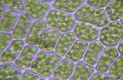
Cell Wall
The materials in a cell wall vary between species, and in plants and fungi also differ between cell types and developmental stages. In plants, the strongest component of the complex cell wall is a carbohydrate called cellulose, which is a polymer of glucose. In bacteria, peptidoglycan forms the cell wall. Archaean cell walls have various compositions, and may be formed of glycoprotein S-layers, pseudopeptidoglycan, or polysaccharides.
Fungi possess cell walls made of the glucosamine polymer chitin, and algae typically possess walls made of glycoproteins and polysaccharides. Unusually, diatoms have a cell wall composed of silicic acid. Often, other accessory molecules are found anchored to the cell wall.
Contents
» Properties
» Rigidity
» Permeability
» Plant cell walls
» Composition
» Formation
» Algal cell walls
» Fungal cell walls
» True fungi
» Fungus-like protists
» Prokaryotic cell walls
» Bacterial cell walls
» Archaeal cell walls
» See also
» References
Properties
Rigidity
The rigidity of cell walls is often over-estimated. In most cells, the cell wall is flexible, meaning that it will bend rather than holding a fixed shape, but has considerable tensile strength. The apparent rigidity of primary plant tissues is a function of hydraulic turgor pressure of the cells and not due to rigid cell walls. This flexibility is seen when plants wilt, so that the stems and leaves begin to droop, or in seaweeds that bend in water currents.The rigidity of healthy plants results from a combination of the wall construction and turgor pressure. As John Howland states it:
“ Think of the cell wall as a wicker basket in which a balloon has been inflated so that it exerts pressure from the inside. Such a basket is very rigid and resistant to mechanical damage. Thus does the prokaryote cell (and eukaryotic cell that possesses a cell wall) gain strength from a flexible plasma membrane pressing against a rigid cell wall. ”
The rigidity of the cell wall thus results in part from inflation of the cell contained. This inflation is a result of the passive uptake of water.
In plants, a secondary cell wall is a thicker additional layer of cellulose which increases wall rigidity. Additional layers may be formed containing lignin in xylem cell walls, or containing suberin in cork cell walls. These compounds are rigid and waterproof, making the secondary wall stiff. Both wood and bark cells of trees have secondary walls. Other parts of plants such as the leaf stalk may acquire similar reinforcement to resist the strain of physical forces.
Certain single-cell protists and algae also produce a rigid wall. Diatoms build a frustule from silica extracted from the surrounding water; radiolarians also produce a test from minerals. Many green algae, such as the Dasycladales encase their cells in a secreted skeleton of calcium carbonate. In each case, the wall is rigid and essentially inorganic.
Permeability
Plant cell walls
Composition
The major carbohydrates making up the primary (growing) plant cell wall are cellulose, hemicellulose and pectin. The cellulose microfibrils are linked via hemicellulosic tethers to form the cellulose-hemicellulose network, which is embedded in the pectin matrix. The most common hemicellulose in the primary cell wall is xyloglucan. In grass cell walls, xyloglucan and pectin are reduced in abundance and partially replaced by glucuronarabinoxylan, a hemicellulose. Primary cell walls characteristically extend (grow) by a mechanism called acid growth, which involves turgor-driven movement of the strong cellulose microfibrils within the weaker hemicellulose/pectin matrix, catalyzed by expansin proteins. The outer part of the primary cell wall of the plant epidermis is usually impregnated with cutin and wax, forming a permeability barrier known as the plant cuticle.Secondary cell walls contain a wide range of additional compounds that modify their mechanical properties and permeability. The major polymers that make up wood (largely secondary cell walls) include cellulose (35 to 50%), xylan, a type of hemicellulose, (20 to 35%) and a complex phenolic polymer called lignin (10 to 25%). Lignin penetrates the spaces in the cell wall between cellulose, hemicellulose and pectin components, driving out water and strengthening the wall. The walls of cork cells in the bark of trees are impregnated with suberin, and suberin also forms the permeability barrier in primary roots known as the Casparian strip. Secondary walls - especially in grasses - may also contain microscopic silica crystals, which may strengthen the wall and protect it from herbivores.
» The middle lamella, a layer rich in pectins. This outermost layer forming the interface between adjacent plant cells and glues them together.
» The primary cell wall, generally a thin, flexible and extensible layer formed while the cell is growing.
» The secondary cell wall, a thick layer formed inside the primary cell wall after the cell is fully grown. It is not found in all cell types. In some cells, such as found xylem, the secondary wall contains lignin, which strengthens and waterpoofs the wall.
Cell walls in some plant tissues also function as storage depots for carbohydrates that can be broken down and resorbed to supply the metabolic and growth needs of the plant. For example, endosperm cell walls in the seeds of cereal grasses, nasturtium, and other species, are rich in glucans and other polysaccharides that are readily digested by enzymes during seed germination to form simple sugars that nourish the growing embryo. Cellulose microfibrils are not readily digested by plants, however.
Formation
In some plants and cell types, after a maximum size or point in development has been reached, a secondary wall is constructed between the plant cell and primary wall. Unlike the primary wall, the microfibrils are aligned mostly in the same direction, and with each additional layer the orientation changes slightly. Cells with secondary cell walls are rigid. Cell to cell communication is possible through pits in the secondary cell wall that allow plasmodesma to connect cells through the secondary cell walls.
Algal cell walls
» Manosyl form microfibrils in the cell walls of a number of marine green algae including those from the genera, Codium, Dasycladus, and Acetabularia as well as in the walls of some red algae, like Porphyra and Bangia.
» Xylanes
» Alginic acid is a common polysaccharide in the cell walls of brown algae
» Sulfonated polysaccharides occur in the cell walls of most algae; those common in red algae include agarose, carrageenan, porphyran, furcelleran and funoran.
Other compounds that may accumulate in algal cell walls include sporopollenin and calcium ions.
The group of algae known as the diatoms synthesize their cell walls (also known as frustules or valves) from silicic acid (specifically orthosilicic acid, H4SiO4). The acid is polymerised intra-cellularly, then the wall is extruded to protect the cell. Significantly, relative to the organic cell walls produced by other groups, silica frustules require less energy to synthesize (approximately 8%), potentially a major saving on the overall cell energy budget and possibly an explanation for higher growth rates in diatoms.
Fungal cell walls
There are several groups of organisms that may be called "fungi". Some of these groups have been transferred out of the Kingdom Fungi, in part because of fundamental biochemical differences in the composition of the cell wall. Most true fungi have a cell wall consisting largely of chitin and other polysaccharides. True fungi do not have cellulose in their cell walls, but some fungus-like organisms do.True fungi
 Chemical structure of a unit from a chitin polymer chain.
» a chitin layer (polymer consisting mainly of unbranched chains of N-acetyl-D-glucosamine)
» a layer of β-1,3-glucan
» a layer of mannoproteins (mannose-containing glycoproteins) which are heavily glycosylated at the outside of the cell.
Fungus-like protists
Prokaryotic cell walls
Bacterial cell walls
Around the outside of the cell membrane is the bacterial cell wall. Bacterial cell walls are made of peptidoglycan (also called murein), which is made from polysaccharide chains cross-linked by unusual peptides containing D-amino acids. Bacterial cell walls are different from the cell walls of plants and fungi which are made of cellulose and chitin, respectively. The cell wall of bacteria is also distinct from that of Archaea, which do not contain peptidoglycan. The cell wall is essential to the survival of many bacteria. The antibiotic penicillin is able to kill bacteria by inhibiting a step in the synthesis of peptidoglycan.There are broadly speaking two different types of cell wall in bacteria, called Gram-positive and Gram-negative. The names originate from the reaction of cells to the Gram stain, a test long-employed for the classification of bacterial species.
Gram-positive bacteria possess a thick cell wall containing many layers of peptidoglycan and teichoic acids. In contrast, Gram-negative bacteria have a relatively thin cell wall consisting of a few layers of peptidoglycan surrounded by a second lipid membrane containing lipopolysaccharides and lipoproteins.

Diagram of a typical gram-negative bacterium, with the thin cell wall sandwiched between the red outer membrane and the thin green plasma membrane
Archaeal cell walls
Although not truly unique, the cell walls of Archaea are unusual. Whereas peptidoglycan is a standard component of all bacterial cell walls, all archaeal cell walls lack peptidoglycan, with the exception of one group of methanogens. In that group, the peptidoglycan is a modified form very different from the kind found in bacteria. There are four types of cell wall currently known among the Archaea.One type of archaeal cell wall is that composed of pseudopeptidoglycan (also called pseudomurein). This type of wall is found in some methanogens, such as Methanobacterium and Methanothermus. While the overall structure of archaeal pseudopeptidoglycan superficially resembles that of bacterial peptidoglycan, there are a number of significant chemical differences. Like the peptidoglycan found in bacterial cell walls, pseudopeptidoglycan consists of polymer chains of glycan cross-linked by short peptide connections.

Schematic of typical gram-positive cell wall showing arrangement of N-Acetylglucosamine and N-Acetlymuramic acid
A second type of archaeal cell wall is found in Methanosarcina and Halococcus. This type of cell wall is composed entirely of a thick layer of polysaccharides, which may be sulfated in the case of Halococcus. Structure in this type of wall is complex and as yet is not fully investigated.
A third type of wall among the Archaea consists of glycoprotein, and occurs in the hyperthermophiles, Halobacterium, and some methanogens. In Halobacterium, the proteins in the wall have a high content of acidic amino acids, giving the wall an overall negative charge. The result is an unstable structure that is stabilized by the presence of large quantities of positive sodium ions that neutralize the charge. Consequently, Halobacterium thrives only under conditions with high salinity.
In other Archaea, such as Methanomicrobium and Desulfurococcus, the wall may be composed only of surface-layer proteins, known as an S-layer. S-layers are common in bacteria, where they serve as either the sole cell-wall component or an outer layer in conjunction with polysaccharides. Most Archaea are Gram-negative, though at least one Gram-positive member is known.
See also
» Plant cellReferences
- Laurence Moire, Alain Schmutz, Antony Buchala, Bin Yan, Ruth E. Stark, and Ulrich Ryser (1999). "Glycerol Is a Suberin Monomer. New Experimental Evidence for an Old Hypothesis". Plant Physiol. 119: 1137-1146
- Buchanan; Gruissem, Jones (2000). Biochemistry & molecular biology of plants (1st ed. ed.). American society of plant physiology.
- Sendbusch, Peter V. (2003-07-31). "Cell Walls of Algae". Botany Online.
- Raven, J. A. (1983). The transport and function of silicon in plants. Biol. Rev. 58, 179-207.
- Furnas, M. J. (1990). "In situ growth rates of marine phytoplankton : Approaches to measurement, community and species growth rates". J. Plankton Res. 12, 1117-1151.
- Hudler, George W. (1998). Magical Mushrooms, Mischievous Molds. Princeton, NJ: Princeton University Press, 7.
- Sengbusch, Peter V. (2003-07-31). "Interactions between Plants and Fungi: the Evolution of their Parasitic and Symbiotic Relations". biologie.uni-hamburg.de.
- Alexopoulos, C. J., C. W. Mims, & M. Blackwell (1996). Introductory Mycology 4. New York: John Wiley & Sons, 687-688.
- Raper, Kenneth B. (1984). The Dictyostelids. Princeton, NJ: Princeton University Press, 99-100.
- van Heijenoort J (2001). "Formation of the glycan chains in the synthesis of bacterial peptidoglycan". Glycobiology 11 (3): 25R – 36R.
- Koch A (2003). "Bacterial wall as target for attack: past, present, and future research". Clin Microbiol Rev 16 (4): 673 – 87.
- Gram, HC (1884). "Über die isolierte F» rbung der Schizomyceten in Schnitt- und Trockenpr» paraten". Fortschr. Med. 2: 185–189.
- Hugenholtz P (2002). "Exploring prokaryotic diversity in the genomic era". Genome Biol 3 (2): REVIEWS0003. doi:10.1186/1471-2148-1-8.
- Walsh F, Amyes S (2004). "Microbiology and drug resistance mechanisms of fully resistant pathogens.". Curr Opin Microbiol 7 (5): 439-44.
- White, David. (1995) The Physiology and Biochemistry of Prokaryotes, pages 6, 12-21. (Oxford: Oxford University Press).
- Brock, Thomas D., Michael T. Madigan, John M. Martinko, & Jack Parker. (1994) Biology of Microorganisms, 7th ed., pages 818-819, 824 (Englewood Cliffs, NJ: Prentice Hall).


