Hormonal Regulation of Calcium Metabolism
 |
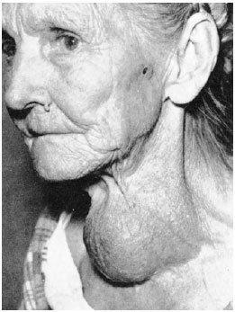
| Figure 36-10 A large goiter caused by iodine deficiency. By enlarging enormously, the thyroid gland can extract enough iodine from the blood to synthesize the body’s requirement for thyroid hormones. |
Closely associated with the thyroid gland and in some animals buried within it are the parathyroid glands. These tiny glands occur as two pairs in humans but vary in number and position in other vertebrates. They were discovered at the end of the nineteenth century when the fatal effects of “thyroidectomy” were traced to unknowing removal of the parathyroid glands together with the thyroid gland. In birds and mammals, including humans, removal of the parathyroid glands causes the level of calcium in the blood to decrease rapidly. This decrease in calcium leads to a serious increase in nervous system excitability, severe muscular spasms and tetany, and finally death. Subsequently, it was discovered that the parathyroid glands secrete a hormone, parathyroid hormone (PTH), which is essential to maintenance of calcium homeostasis. Calcium ions are extremely important for formation of healthy bones. In addition, they are needed for neurotransmitter and hormone release, for muscle contraction, and for blood clotting.
Before considering how hormones maintain calcium homeostasis, it is helpful to summarize mineral metabolism in bone, a densely packed storehouse of both calcium and phosphorus. Bone contains approximately 98% of the calcium and 80% of the phosphorus in humans. Although bone is second only to teeth as the most durable material in the body (as evidenced by survival of fossil bones for millions of years) it is in a state of constant turnover in living vertebrates. Bone-building cells (osteoblasts) synthesize organic fibers of bone matrix which later become mineralized with a form of calcium phosphate called hydroxyapatite. Bone-resorbing cells (osteoclasts) are giant cells that dissolve the bony matrix, releasing calcium and phosphate into the blood. These opposing activities allow bone constantly to remodel itself, especially in a growing animal, for structural improvements to counter new mechanical stresses on the body. They additionally provide a vast and accessible reservoir of minerals that can be withdrawn as needed for general cellular requirements.
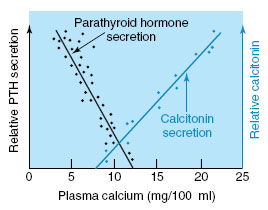
The level of calcium in the blood is maintained by three hormones that coordinate the absorption, storage, and excretion of calcium ions. If blood calcium should decrease slightly, the parathyroid gland increases its secretion of PTH. This increase stimulates osteoclasts to dissolve bone adjacent to these cells, thus releasing calcium and phosphate into the bloodstream and returning blood calcium level to normal. PTH also decreases the rate of calcium excretion by the kidney and increases production of the hormone 1,25-dihydroxyvitamin D (see the following text). PTH levels vary inversely with blood calcium level, as shown in Figure 36-11.
 |
| Figure 36-11 How rate of secretion of parathyroid hormone (PTH) and calcitonin respond to changes in blood calcium level in a mammal. |
A second hormone involved in calcium metabolism in all tetrapods is derived from vitamin D. Vitamin D, like all vitamins, is a dietary requirement. But unlike other vitamins, vitamin D may also be synthesized in the skin from a precursor by irradiation with ultraviolet light from the sun. Vitamin D is then converted in a two-step oxidation to a hormonal form, 1,25- dihydroxyvitamin D. This steroid hormone is essential for active calcium absorption by the gut (Figure 36-12). Production of 1,25-dihydroxyvitamin D is stimulated by low plasma phosphate as well as by an increase in PTH secretion.
In humans, a deficiency of vitamin D causes rickets, a disease characterized by low blood calcium and weak, poorly calcified bones that tend to bend under postural and gravitational stresses. Rickets has been called a disease of northern winters, when sunlight is minimal. It once was common in the smoke-darkened cities of England and Europe.
A third calcium-regulating hormone, calcitonin, is secreted by specialized cells (C cells) in the thyroid gland of mammals and in the ultimobranchial gland of other vertebrates. Calcitonin is released in response to elevated levels of calcium in the blood. It rapidly suppresses calcium withdrawal from bone, decreases intestinal absorption of calcium, and increases excretion of calcium by the kidneys. Calcitonin therefore protects the body against an increase in level of calcium in the blood, just as parathyroid hormone protects it from a decrease in blood calcium (Figure 36-12). Calcitonin has been identified in all vertebrate groups, but its importance is uncertain because replacement of calcitonin is not required for maintenance of calcium homeostasis, at least in humans, if the thyroid gland is surgically removed (also removing the C cells).


