Phylum Ectoprocta (Bryozoa)
Phylum Ectoprocta
(Bryozoa)
Ectoprocta (ek-to-prok´ta) (Gr. ektos, outside, + proktos, anus) have long been called bryozoans, or moss animals (Gr. bryon, moss, + zoon, animal), a term that originally included Entoprocta also. However, because entoprocts are pseudocoelomates and have the anus located within the tentacular crown, they are commonly separated from ectoprocts, which, like other lophophorates, are eucoelomate and have the anus outside the circle of tentacles. Some authors continue to use the name “Bryozoa” but exclude entoprocts from the group.
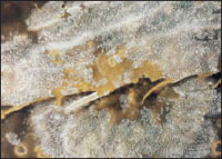
Of the 4000 or so species of ectoprocts, few are more than 0.5 mm long. All are aquatic, both freshwater and marine, but are found largely in shallow waters. With very few exceptions they are colony builders. Ectoprocts have become diverse and abundant. They have left a rich fossil record since the Ordovician period. Marine forms today exploit all kinds of firm surfaces, such as shells, rocks, large brown algae, mangrove roots, and ship bottoms.
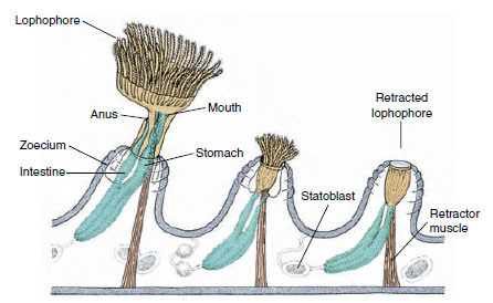
Each member of a colony lives in a tiny chamber, called a zoecium, which is secreted by its epidermis (Figure 22-2). Each individual, or zooid, consists of a feeding polypide and a case-forming cystid. The polypide includes the lophophore, digestive tract, muscles, and nerve centers. The cystid is the body wall of the animal, together with its secreted exoskeleton. The exoskeleton, or zoecium, may, according to species, be gelatinous, chitinous, or stiffened with calcium and possibly also impregnated with sand. The shape may be boxlike, vaselike, oval, or tubular.
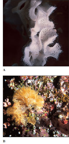
Some colonies form limy encrustations on seaweed, shells, and rocks; others form fuzzy or shrubby growths on erect, branching colonies that look like seaweed (Figure 22-3). Some ectoprocts might easily be mistaken for hydroids but can be distinguished under a microscope by the presence of an anus (Figure 22-2). In some freshwater forms individuals are borne on finely branching stolons that form delicate tracings on the underside of rocks or plants. Other freshwater
ectoprocts are embedded in
large masses of gelatinous material.
Although zooids are minute, the
colonies may be several centimeters
in diameter, some encrusting colonies
may be a meter or more in width (Figure
22-4), and erect forms may reach
30 cm or more in height. Freshwater
ectoprocts may form mosslike colonies
on stems of plants or on rocks, usually
in shallow ponds or pools. They may be
able to slide along slowly on the object
that supports them.
Polypides live a type of jack-inthe- box existence, popping up to feed and then quickly withdrawing into their little chamber, which often has a tiny trapdoor (operculum) that shuts to conceal its inhabitant. To extend the tentacular crown, certain muscles contract, which increases the hydrostatic pressure within the body cavity and pushes the lophophore out by a hydraulic mechanism. Other muscles can contract to withdraw the crown to safety with great speed.
The lophophore ridge tends to be circular in marine ectoprocts (Figure 22-5A) and U-shaped in freshwater species (Figure 22-5B). When feeding, the animal extends the lophophore and spreads the tentacles out into a funnel. Cilia on the tentacles draw water into the funnel and out between the tentacles. Food particles caught by cilia in the funnel are drawn into the mouth, both by a pumping action of the muscular pharynx and by action of cilia in the pharynx. Undesirable particles can be rejected by reversing the ciliary action, by drawing the tentacles close together, or by retracting the whole lophophore into the zoecium. Digestion in the ciliated, U-shaped digestive tract appears to be extracellular for protein and starches and intracellular for fats.
Respiratory, vascular, and excretory organs are absent. Gaseous exchange is through the body surface, and since ectoprocts are small, coelomic fluid is adequate for internal transport. Coelomocytes engulf and store waste materials. A ganglionic mass and a nerve ring surround the pharynx, but no sense organs are present. A septum divides the mesocoel in the lophophore from the larger posterior metacoel. A protocoel and epistome occur only in freshwater ectoprocts. Pores in the walls between adjoining zooids permit exchange of materials by way of the coelomic fluid.
Most colonies contain only feeding individuals, but polymorphism occurs in some species. One type of modified zooid resembles a bird beak that snaps at small invading organisms that might foul a colony. Another type has a long bristle that sweeps away foreign particles.
Most ectoprocts are hermaphroditic. Some species shed eggs into seawater, but most brood their eggs, some within the coelom and some externally in a special ovicell, which is a modified zoecium in which an embryo develops. Cleavage is radial but apparently mosaic. Little is known of mesoderm derivation. Larvae of nonbrooding species have a functional gut and swim about for a few months before settling; larvae of brooding species do not feed and settle after a brief free-swimming existence. They attach to the substratum by mucopolysaccharide and protein secretions from an adhesive sac, then metamorphose to the adult form.
Ectoprocts reproduce asexually by budding and form colonies. Freshwater ectoprocts have another type of budding that produces statoblasts (Figure 22-6), which are hard, resistant capsules containing a mass of germinative cells. Statoblasts are formed during the summer and fall. When the colony dies in late autumn, the statoblasts remain, and in spring they give rise to new polypides and eventually to new colonies.
Ectoprocta (ek-to-prok´ta) (Gr. ektos, outside, + proktos, anus) have long been called bryozoans, or moss animals (Gr. bryon, moss, + zoon, animal), a term that originally included Entoprocta also. However, because entoprocts are pseudocoelomates and have the anus located within the tentacular crown, they are commonly separated from ectoprocts, which, like other lophophorates, are eucoelomate and have the anus outside the circle of tentacles. Some authors continue to use the name “Bryozoa” but exclude entoprocts from the group.
Of the 4000 or so species of ectoprocts, few are more than 0.5 mm long. All are aquatic, both freshwater and marine, but are found largely in shallow waters. With very few exceptions they are colony builders. Ectoprocts have become diverse and abundant. They have left a rich fossil record since the Ordovician period. Marine forms today exploit all kinds of firm surfaces, such as shells, rocks, large brown algae, mangrove roots, and ship bottoms.
Each member of a colony lives in a tiny chamber, called a zoecium, which is secreted by its epidermis (Figure 22-2). Each individual, or zooid, consists of a feeding polypide and a case-forming cystid. The polypide includes the lophophore, digestive tract, muscles, and nerve centers. The cystid is the body wall of the animal, together with its secreted exoskeleton. The exoskeleton, or zoecium, may, according to species, be gelatinous, chitinous, or stiffened with calcium and possibly also impregnated with sand. The shape may be boxlike, vaselike, oval, or tubular.
 |
| Figure 22-2 Small portion of freshwater colony of Plumatella (phylum Ectoprocta), which grows on the underside of rocks. These tiny individuals disappear into their chitinous zoecia when disturbed. Statoblasts are resistant capsules containing germinative cells. |
 |
| Figure 22-3 Colonies of marine ectoprocts. A, The zooids are extended in this lacy colony of Triphyllozoon sp. B, Reteporella graffei has upright, branching colonies. |
Some colonies form limy encrustations on seaweed, shells, and rocks; others form fuzzy or shrubby growths on erect, branching colonies that look like seaweed (Figure 22-3). Some ectoprocts might easily be mistaken for hydroids but can be distinguished under a microscope by the presence of an anus (Figure 22-2). In some freshwater forms individuals are borne on finely branching stolons that form delicate tracings on the underside of rocks or plants. Other freshwater
 |
| Figure 22-4 Skeletal remains of a colony of Membranipora, a marine encrusting form of Ectoprocta. Each little oblong zoecium is the calcareous former home of a tiny ectoproct. |
Polypides live a type of jack-inthe- box existence, popping up to feed and then quickly withdrawing into their little chamber, which often has a tiny trapdoor (operculum) that shuts to conceal its inhabitant. To extend the tentacular crown, certain muscles contract, which increases the hydrostatic pressure within the body cavity and pushes the lophophore out by a hydraulic mechanism. Other muscles can contract to withdraw the crown to safety with great speed.
The lophophore ridge tends to be circular in marine ectoprocts (Figure 22-5A) and U-shaped in freshwater species (Figure 22-5B). When feeding, the animal extends the lophophore and spreads the tentacles out into a funnel. Cilia on the tentacles draw water into the funnel and out between the tentacles. Food particles caught by cilia in the funnel are drawn into the mouth, both by a pumping action of the muscular pharynx and by action of cilia in the pharynx. Undesirable particles can be rejected by reversing the ciliary action, by drawing the tentacles close together, or by retracting the whole lophophore into the zoecium. Digestion in the ciliated, U-shaped digestive tract appears to be extracellular for protein and starches and intracellular for fats.
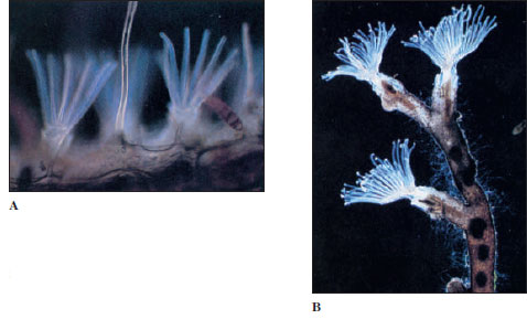 |
| Figure 22-5 A, Ciliated lophophore of Electra pilosa, a marine ectoproct. B, Plumatella repens, a freshwater bryozoan (phylum Ectoprocta). It grows on the underside of rocks and vegetation in lakes, ponds, and streams. |
Respiratory, vascular, and excretory organs are absent. Gaseous exchange is through the body surface, and since ectoprocts are small, coelomic fluid is adequate for internal transport. Coelomocytes engulf and store waste materials. A ganglionic mass and a nerve ring surround the pharynx, but no sense organs are present. A septum divides the mesocoel in the lophophore from the larger posterior metacoel. A protocoel and epistome occur only in freshwater ectoprocts. Pores in the walls between adjoining zooids permit exchange of materials by way of the coelomic fluid.
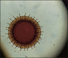 |
| Figure 22-6 Statoblast of a freshwater ectoproct Cristatella. This statoblast is about 1 mm in diameter and bears hooked spines. |
Most colonies contain only feeding individuals, but polymorphism occurs in some species. One type of modified zooid resembles a bird beak that snaps at small invading organisms that might foul a colony. Another type has a long bristle that sweeps away foreign particles.
Most ectoprocts are hermaphroditic. Some species shed eggs into seawater, but most brood their eggs, some within the coelom and some externally in a special ovicell, which is a modified zoecium in which an embryo develops. Cleavage is radial but apparently mosaic. Little is known of mesoderm derivation. Larvae of nonbrooding species have a functional gut and swim about for a few months before settling; larvae of brooding species do not feed and settle after a brief free-swimming existence. They attach to the substratum by mucopolysaccharide and protein secretions from an adhesive sac, then metamorphose to the adult form.
Ectoprocts reproduce asexually by budding and form colonies. Freshwater ectoprocts have another type of budding that produces statoblasts (Figure 22-6), which are hard, resistant capsules containing a mass of germinative cells. Statoblasts are formed during the summer and fall. When the colony dies in late autumn, the statoblasts remain, and in spring they give rise to new polypides and eventually to new colonies.
Support our developers


