The Many Forms of Cellulose — A Brief Introduction to the Structure and Different Crystalline Forms of Cellulose
Unlike most known biopolymers, cellulose is a simple molecule that consists of an
assembly of β-1,4-linked glucan chains. As a result, cellulose is defined less by its
primary structure (β-1,4-linked glucose residues with cellobiose being the repeating
unit in all chains) and more by its secondary and higher-order structure in
which the chains interact via intramolecular and intermolecular hydrogen bonds,
as well as van der Waals interactions, to give rise to different forms of cellulose
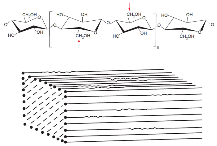
(Fig. 6.1) (O'Sullivan, 1997). Cellulose exhibits polymorphism, and the different
forms of cellulose are usually defined by their crystalline forms, although reference
is also made to other forms of cellulose such as noncrystalline cellulose,
amorphous cellulose, and more recently nematic-ordered cellulose (Kondo
et al.,
2001). Whereas, the glucan chains are arranged in a specific manner with respect
to each other in crystalline cellulose, no specific arrangement of the glucan chains
occur in noncrystalline or amorphous cellulose. In contrast, nematic-ordered
cellulose is highly ordered but not crystalline and is obtained by uniaxial
stretching of water-swollen cellulose (Kondo
et al., 2004).
 |
| FIGURE 6.1 Top image is the structural formula for the β-1,4-linked glucan chain of cellulose.
The bracketed region indicates the basic repeat unit, cellobiose, in the chain. The glucan chain has a
twofold symmetry. The bottom image is a schematic representation of a crystalline cellulose I
microfibril. (Reproduced from Brown, Jr. R. M., J. Poly. Sci. Part A Poly. Chem. 42, 489–495.) |
 |
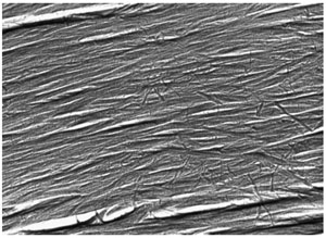
| FIGURE 6.2 Freeze fracture image of cellulose
microfibrils in the secondary wall of a developing
cotton fiber. (Unpublished image from
R.
Malcolm Brown, Jr. and Kazuo Okuda.) |
In general, cellulose produced by living organisms occurs as cellulose I and is
assembled in a structure referred to as a microfibril (Fig. 6.2). The properties of the
microfibril are determined by its size, shape, and crystallinity. The glucan chains
in cellulose I are arranged in a parallel manner, and depending upon the arrangement
of these chains, two crystalline forms of cellulose I—I
α and I
β—have been identified (Attala and Vanderhart, 1984). The more thermodynamically stable
form of cellulose is cellulose II, and in this allomorph the glucan chains are
arranged in an antiparallel manner. Cellulose II is produced in nature by certain
organisms or under specific conditions but is generally obtained by an irreversible process upon chemical treatment (mercerization or solubilization) of native
cellulose I. Furthermore, cellulose IIII and cellulose IIIII are obtained from cellulose
I and cellulose II, respectively, in a reversible process, by treatment with
liquid ammonia or some amines and the subsequent evaporation of excess ammonia,
and cellulose IVI and cellulose IVII are obtained irreversibly by heating
cellulose IIII and cellulose IIIII respectively to 206 °C in glycerol (O'Sullivan,
1997). Implicit in the biosynthesis of cellulose is the role of the cellulosesynthesizing
machinery that allows synthesis and organization of a metastable
form of cellulose (cellulose I) that is found to be desirable in living organisms in
comparison to the more stable cellulose II product. Whereas the assembly of the
glucan chains (crystallization) endows cellulose with its characteristic properties,
it is the synthesis of these β-1,4-linked glucan chains (polymerization) that is the
focus of research for most biologists.


