Phylum Acanthocephala
Phylum
Acanthocephala
Members of phylum Acanthocephala (a-kan´tho-sef´a-la) (Gr. akantha, spine or thorn, + kephale, head) are commonly known as “spiny-headed worms.” The phylum derives its name from one of its most distinctive features, a cylindrical, invaginable proboscis bearing rows of recurved spines, by which it attaches itself to the intestine of its host. All acanthocephalans are endoparasitic, living as adults in the intestine of vertebrates.
Various species range in size from less than 2 mm to more than 1 m in length, with females of a species usually larger than males. The body is usually bilaterally flattened, with numerous transverse wrinkles. Worms are typically cream color but may be yellowish or brown as a result of absorption of pigments from the intestinal contents.
Acanthocephalans inflict traumatic damage by penetrating the host’s intestinal wall with the spiny proboscis. In many cases there is remarkably little inflammation, but in some species the inflammatory response of the host is intense. Infection with these worms can cause great pain, particularly if the gut wall is completely perforated.
The phylum is cosmopolitan, and more than 500 species are known, most of which parasitize fish, birds, and mammals. However, no species is normally a parasite of humans, although species that usually occur in other hosts occasionally infect humans. Macracanthorhynchus hirudinaceus (Gr. makros, long, large, + akantha, spine, thorn, + rhynchos, beak) occurs throughout the world in the small intestine of pigs and sometimes in other mammals.
Larvae of spiny-headed worms develop in arthropods, either crustaceans or insects, depending on the species.
Form and Function
In life the body is somewhat flattened,
although it is usual for specimens to be
treated with tap water before fixation
so that fixed specimens are turgid and
cylindrical (Figure 15-19C).
The body wall is syncytial, and its surface is punctured by minute crypts 4 to 6 µm deep, which greatly increase the surface area of the tegument. About 80% of the thickness of the tegument is the radial fiber zone, which contains a lacunar system of ramifying fluid-filled canals (Figure 15-19A and B). Curiously, body-wall muscles are tubelike and filled with fluid. The tubes in the muscles are continuous with the lacunar system; therefore circulation of lacunar fluid may well bring nutrients to and remove wastes from the muscles. There is no heart or other circulatory system, and contraction of the muscles would serve to move lacunar fluid through the canals and muscles. Both longitudinal and circular body-wall muscles are present.
The proboscis, which bears rows of recurved hooks, is attached to the neck region (Figure 15-19) and can be inverted into a proboscis receptacle by retractor muscles. Attached to the neck region (but not within the proboscis) are two elongated lemnisci (extensions of the tegument and lacunar system) that may serve as reservoirs of lacunar fluid from the proboscis when that organ is invaginated.
There is no respiratory system. When present, the excretory system consists of a pair of protonephridia with flame cells. These unite to form a common tube opening into the sperm duct or uterus.
The nervous system has a central ganglion within the proboscis receptacle and nerves to the proboscis and body. There are sensory endings on the proboscis and genital bursa.
Acanthocephalans have no digestive tract, and they must absorb all nutrients through their tegument. They can absorb various molecules by specific membrane transport mechanisms, and other substances can cross the tegumental membrane by pinocytosis (probably potocytosis). The tegument bears some enzymes, such as peptidases, which can cleave several dipeptides, and the amino acids are then absorbed by the worm. Like cestodes, acanthocephalans require host dietary carbohydrate, but their mechanism for absorption of glucose is different. As glucose is absorbed, it is rapidly phosphorylated and compartmentalized, so that a metabolic “sink” is created into which glucose from the surrounding medium can flow. Glucose diffuses down the concentration gradient into the worm because it is constantly removed as soon as it enters.
Acanthocephalans are dioecious. A pair of tubular genital ligaments, or ligament sacs, extends posteriorly from the end of the proboscis receptacle. Males have a pair of testes, each with a vas deferens, and a common ejaculatory duct that ends in a small penis. During copulation sperm are ejected into the vagina, travel up the genital duct, and escape into the pseudocoel.
In females the ovarian tissue in the
ligament sac breaks up into ovarian
balls that rupture the ligament sacs and
float free in the pseudocoel. One of the
ligament sacs leads to a funnel-shaped uterine bell that receives the developing
shelled embryos and passes them
on to the uterus (Figure 15-20). An
interesting and unique selective apparatus
operates here. Fully developed
embryos are slightly longer than immature
ones, and they are passed on into
the uterus, while immature eggs are
retained for further maturation.
The shelled embryos, which are discharged in the feces of the vertebrate host, do not hatch until eaten by an intermediate host. For M. hirudinaceus this is any of several species of soil-inhabiting beetle larvae, especially scarabeids. Grubs of the June beetle (Phyllophaga) are frequent hosts. Here the larva (acanthor) burrows through the intestine and develops into a juvenile (cystacanth) in the insect’s hemocoel. Pigs become infected by eating the grubs. Multiple infections may do considerable damage to a pig’s intestine, and perforations can occur.
Members of phylum Acanthocephala (a-kan´tho-sef´a-la) (Gr. akantha, spine or thorn, + kephale, head) are commonly known as “spiny-headed worms.” The phylum derives its name from one of its most distinctive features, a cylindrical, invaginable proboscis bearing rows of recurved spines, by which it attaches itself to the intestine of its host. All acanthocephalans are endoparasitic, living as adults in the intestine of vertebrates.
Various species range in size from less than 2 mm to more than 1 m in length, with females of a species usually larger than males. The body is usually bilaterally flattened, with numerous transverse wrinkles. Worms are typically cream color but may be yellowish or brown as a result of absorption of pigments from the intestinal contents.
Acanthocephalans inflict traumatic damage by penetrating the host’s intestinal wall with the spiny proboscis. In many cases there is remarkably little inflammation, but in some species the inflammatory response of the host is intense. Infection with these worms can cause great pain, particularly if the gut wall is completely perforated.
The phylum is cosmopolitan, and more than 500 species are known, most of which parasitize fish, birds, and mammals. However, no species is normally a parasite of humans, although species that usually occur in other hosts occasionally infect humans. Macracanthorhynchus hirudinaceus (Gr. makros, long, large, + akantha, spine, thorn, + rhynchos, beak) occurs throughout the world in the small intestine of pigs and sometimes in other mammals.
Larvae of spiny-headed worms develop in arthropods, either crustaceans or insects, depending on the species.
Form and Function
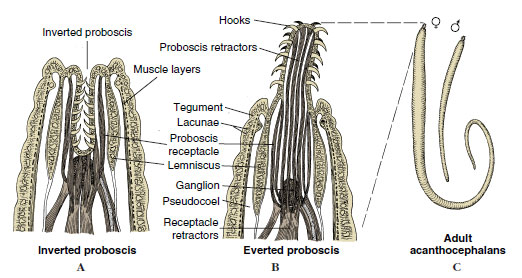 |
| Figure 15-19 Structure of a spiny-headed worm (phylum Acanthocephala). A and B, Eversible spiny proboscis by which the parasite attaches to the intestine of its host, often doing great damage. Because they lack a digestive tract, food is absorbed through the tegument. C, Male is typically smaller than female. |
The body wall is syncytial, and its surface is punctured by minute crypts 4 to 6 µm deep, which greatly increase the surface area of the tegument. About 80% of the thickness of the tegument is the radial fiber zone, which contains a lacunar system of ramifying fluid-filled canals (Figure 15-19A and B). Curiously, body-wall muscles are tubelike and filled with fluid. The tubes in the muscles are continuous with the lacunar system; therefore circulation of lacunar fluid may well bring nutrients to and remove wastes from the muscles. There is no heart or other circulatory system, and contraction of the muscles would serve to move lacunar fluid through the canals and muscles. Both longitudinal and circular body-wall muscles are present.
The proboscis, which bears rows of recurved hooks, is attached to the neck region (Figure 15-19) and can be inverted into a proboscis receptacle by retractor muscles. Attached to the neck region (but not within the proboscis) are two elongated lemnisci (extensions of the tegument and lacunar system) that may serve as reservoirs of lacunar fluid from the proboscis when that organ is invaginated.
There is no respiratory system. When present, the excretory system consists of a pair of protonephridia with flame cells. These unite to form a common tube opening into the sperm duct or uterus.
The nervous system has a central ganglion within the proboscis receptacle and nerves to the proboscis and body. There are sensory endings on the proboscis and genital bursa.
Acanthocephalans have no digestive tract, and they must absorb all nutrients through their tegument. They can absorb various molecules by specific membrane transport mechanisms, and other substances can cross the tegumental membrane by pinocytosis (probably potocytosis). The tegument bears some enzymes, such as peptidases, which can cleave several dipeptides, and the amino acids are then absorbed by the worm. Like cestodes, acanthocephalans require host dietary carbohydrate, but their mechanism for absorption of glucose is different. As glucose is absorbed, it is rapidly phosphorylated and compartmentalized, so that a metabolic “sink” is created into which glucose from the surrounding medium can flow. Glucose diffuses down the concentration gradient into the worm because it is constantly removed as soon as it enters.
Acanthocephalans are dioecious. A pair of tubular genital ligaments, or ligament sacs, extends posteriorly from the end of the proboscis receptacle. Males have a pair of testes, each with a vas deferens, and a common ejaculatory duct that ends in a small penis. During copulation sperm are ejected into the vagina, travel up the genital duct, and escape into the pseudocoel.
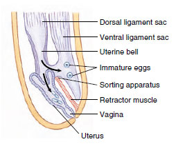 |
| Figure 15-20 Scheme of the genital selective apparatus of a female acanthocephalan. It is a unique device for separating immature from mature fertilized eggs. Eggs containing larvae enter the terine bell and pass on to the uterus and exterior. Immature eggs are shunted into the ventral ligament sac or into the pseudocoel to undergo further development. |
The shelled embryos, which are discharged in the feces of the vertebrate host, do not hatch until eaten by an intermediate host. For M. hirudinaceus this is any of several species of soil-inhabiting beetle larvae, especially scarabeids. Grubs of the June beetle (Phyllophaga) are frequent hosts. Here the larva (acanthor) burrows through the intestine and develops into a juvenile (cystacanth) in the insect’s hemocoel. Pigs become infected by eating the grubs. Multiple infections may do considerable damage to a pig’s intestine, and perforations can occur.
Support our developers


