Hanging-Drop and Wet-Mount Oreparations
Now that you have been oriented to some basic tools and methods used in microbiology, we shall begin our study of microorganisms by learning how to make preparations to study their morphology under the microscope.The simplest method for examining living microorganisms is to suspend them in a fluid (water, saline, or broth) and prepare either a “hanging drop” or a simple “wet mount.”The slide for a hanging drop is ground with a concave well in the center; the cover glass holds a drop of the suspension. When the cover glass is inverted over the well of the slide, the drop hangs from the glass in the hollow concavity of the slide (fig. 3.1, step 4).
Microscopic study of such a wet preparation can provide useful information. Primarily, the method is used to determine whether or not an organism is motile, but it also permits an undistorted view of natural patterns of cell groupings andof individual cell shape. Hanging-drop preparations can be observed for a fairly long time, because the drop does not dry up quickly. Wet-mounted preparations are used primarily to detect microbial motility rapidly. The fluid film is thinner than that of hanging-drop preparations and therefore the preparation tends to dry up more quickly, even when sealed. Although the hanging drop is the
classical method for viewing unstained microorganisms, the wet mount is easier to perform and usually provides sufficient information.
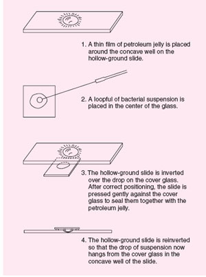 |
| Figure 3.1 Hanging-drop preparation using petroleum jelly to seal the cover glass to the slide. |
Support our developers


