Euglenophyta
FIGURE 2.86 Ejected mucocyst of Peranema trichophorum.
Trichocysts are present only in some phagotrophic species, such as
Entosiphon and
Peranema trichophorum. In Peranema ejectile organelles or mucocysts are located beneath the cell surface, often subtending the pellicular articulations. In transverse sections they appear as hollow tubes of amorphous material, with low electron density, enclosed within a vesicle bounded by single membrane, often found near the Golgi apparatus; or they show a content of greater density, and may subtend the pellicle or protrude through it. Mucocysts subtending the pellicular articulations are ejected from the cell through the grooves. They are characterized by three distinct regions: an inner tube 1.20 mm long and 1.18 mm wide with helical striations; an outer tube 0.77 mm long and 0.21 mm wide with diamond-shaped pattern; and a dense middle band 0.07 mm in width. Often a crown of dense fibrillar material occurs at the tips of the mucocyst (Figure 2.86). Small slimeextruding mucus bodies are the trichocyst counterparts commonly found in the non-phagotrophs.
FIGURE 2.86 Ejected mucocyst of Peranema trichophorum.
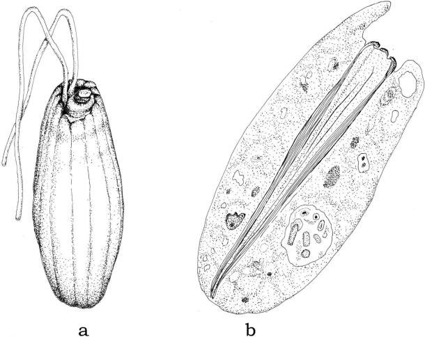
FIGURE 2.87 Cell of Entosiphon sp. (a); longitudinal section of the cell, showing the structure of the feeding apparatus (b).
A feeding apparatus is present in many Euglenophyceae, such as
Diplonema,
Ploeotia, or
Entosiphon. In the latter (Figure 2.87a), it consists of a cytostome surrounded by four centrally located vanes (series of plicate folds or curved ribbons) and supported by rods composed of closely packed microtubules. Near the base of the feeding apparatus, one of the rods splits
giving rise to a total of three stout rods that extend nearly the length of the cell. About one third the distance down the apparatus (from the anterior end) the number of microtubules per rod increases dramatically and then decreases in number towards the base giving the apparatus the overall appearance of a cone with one side open (Figure 2.87b). This feeding apparatus can be moved in and out, extending toward the anterior of the cell and then withdrawing down into the cell for a distance of 3–5 mm. This motion allows food to be drawn into the cell.
FIGURE 2.87 Cell of Entosiphon sp. (a); longitudinal section of the cell, showing the structure of the feeding apparatus (b).
Chlorarachniophyta
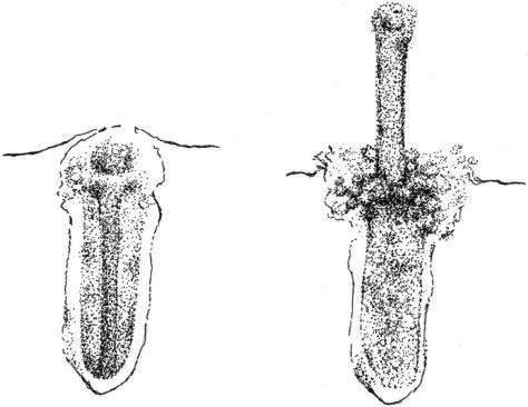
FIGURE 2.88 Ejectile organelle of Bigelowiella natans before (left) and during discharge (right).
Structures interpreted as ejectile organelles (extrusomes) are commonly seen in
Bigelowiella natans. Each organelle resides in a vesicle and consists of two parts. The anterior part is very short, measuring ca. 0.15 µm in length, whereas the posterior part is ca. 0.55 µm long. The posterior part has a very distinct substructure, comprising a core surrounded by a less opaque cylinder. The cylinder is lined by a thin membrane-like structure, whereas the core comprises a thinner membranous structure surrounded by a more opaque one. The structure of the anterior part of the ejectile organelle is less well defined. Although it is clear that the membrane of the surrounding vesicle forms an indentation ring that separates the two parts of the ejectile organelle, the path of the membrane in this area is less clear. It appears that only the core of the organelle is discharged, the core apparently turning inside out during discharge. The surrounding cylinder seems to remain in place. The fate of the anterior part of the ejectile organelle after discharge has not been ascertained (Figure 2.88).
FIGURE 2.88 Ejectile organelle of Bigelowiella natans before (left) and during discharge (right).
ChlorophytaSome
Pyramimonas species (Prasinophyceae) contain ejectile structures similar to the ejectosomes of Cryptophyta. In the undischarged state, they consist of a coiled ribbon, which upon discharge forms a hollow tube tapered at both ends.


