Other Intraflagellar Accessory Structures
Other intraflagellar accessory structures are present in dinoflagellates besides the PFR, the so-called R fiber (Rf), and the so-called striated fiber (Sf). These structures do not show any kind of lattice or precisely organized structure. However, they deserve mentioning because they run for long distances along the axonemal structure and therefore are also candidate for modulating the axonemal beating, possibly by Ca2
+-dependent contraction or through dipole–dipole forces.
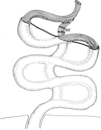
FIGURE 2.38 Schematic drawing of longitudinal flagellum of Ceratium furca during its contraction showing the striated Rf.
The Rf is made of thin filaments 2–4 nm in diameter. It may be as large as the axoneme or the
PFR in diameter (300–500 nm), runs along the major part of the axoneme and is attached to it via
the PFR, the PFR linking the Rf to the axoneme. The Rf may contract and shows transversal striations
of variable periodicities and thickness only during its contraction, but not in its relaxed or fully contracted state (Figure 2.38). The precise structure of the Rf varies according to the fixation conditions for TEM, mostly depending on the Ca2
+ concentration, suggesting that its contractility is Ca2
+-dependent. In some dinoflagellates such as Ceratium furca, the Rf is a good candidate for the induction of the complete retraction of the longitudinal flagellum in the flagellar pocket, a movement that cannot be explained by the axoneme structure itself. In this retracted state, the Rf is contracted and the axoneme is highly folded (more tightly than during the usual flagellar beating). Therefore, the Rf could modulate the properties of the PFR and of axoneme motility through constraints imposed to the PFR.
FIGURE 2.38 Schematic drawing of longitudinal flagellum of Ceratium furca during its contraction showing the striated Rf.
The so-called Sf is also made of thin filaments. It is much smaller than the PFR or the axoneme
in diameter (about 35 nm), and runs along three fourths of the axoneme. Its transversal striations
suggest its implication in contractile processes that could modulate axonemal motility through
gradual changes in the axonemal wavelength or amplitude.


