Transition Zone
While both the flagellar axoneme and the basal body that continues it are very constant in morphology among the different algae, the transition region where they meet varies considerably,
and is considered one of the most useful indicators of phylogenetic relationships. This region sometimes contains particular structures such as helices, star-shaped bodies, and transverse partitions
referred to as basal plates. Five main types of transition zones may be distinguished, taking into
account that secondary variations can appear within each.
Type 1 (Figure 2.39) appears the simplest, with only one basal plate situated at the level of the
point of inflexion of the flagellar membrane. Immediately above it, the flagellum shows a slight narrowing. Radial fibers connect the peripheral doublets with submembrane swellings. This type of
transition zone is present in the Phaeophyceae (Heterokontophyta).
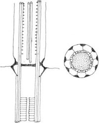
FIGURE 2.39 Type 1 transition zone (Phaeophyceae, Heterokontophyta).
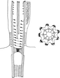
FIGURE 2.40 Type 2 transition zone (Euglenophyta).
In Type 2 (Figure 2.40) the basal plate is replaced by a sort of plug located beneath the point of
inflexion of the flagellar membrane, far from the point of origin of the central pair of doublets. In
this transition zone the nine outer doublets show a strong dilatation, which leaves room for a fibrillar spiral suspended on the doublets by short spokes. The spiral can reach the central doublet, or be much shorter. Each outer doublet is associated via other spokes with a thickening of the membrane, whose folding forms a star with nine characteristics arms (Figure 2.41a and 2.41b). This transition region is typical of the Euglenophyta. Among these algae, Entosiphon sulcatum is unique for its long transition region, the spiral of which surrounds the proximal 1 µm of the central doublet.
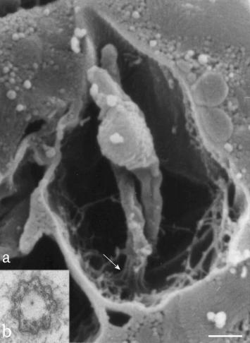
FIGURE 2.41 Scanning electron microscopy image of the reservoir region of Euglena gracilis, showing the two flagella arising from the bottom of the reservoir. The arrow points to the thickened folding of the flagellum membrane, typical of the transition zone (a). Transmission electron microscopy image of the transition zone in transverse section, showing the characteristic star configuration (b). (Bar: 0.50 µm.)
Type 3 transition regions possess a double system of complex plates. Variations exist due to the distance separating the plates or to the presence of interposition material between them. In Dinophyta, the basal plate is duplicated and ring-shaped; above this there are two median discs, the uppermost supporting the central doublet of the axoneme, (Figure 2.42a). In Cryptophyta, an apical plate is located at the level of the narrowing of the flagellum above the point of inflexion of the flagellar membrane; the central doublet is right above this plate. A second plate bearing a ring-shaped thickening is located beneath it. In the Glaucophyta of the genus Cyanophora the apical plate has the same localization present in Cryptophyta, but it is ring-shaped and traversed by the central doublet that continues towards the basal plate situated at the level of the point of inflexion of the flagellar membrane (Figure 2.42b). Further variations of this type are present in Haptophyta, where two widely spaced plates are present, the apical below the central doublet. Each plate corresponds to a flagellar constriction, and the space between them possibly contains fibrillar material; in cross-section a stellate structure is visible, the arm of which connects with the a-tubules of the peripheral doublets (Figure 2.42c).
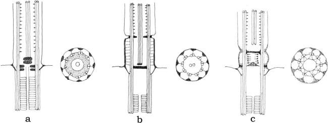
FIGURE 2.42 Type 3 transition zone of Dinophyta (a), Glaucophyta (b), and Haptophyta (c).
In Type 4 (Figure 2.43) there is only one basal plate situated at the point where the flagellum emerges, but this type of transition region is characterized by a very peculiar structure called “transitional helix.” In longitudinal section this appears as a double row of punctae equidistant from the doublets, representing the four to six turns of a helix (Figure 2.44a and b). Some variations occur in the number of gyres, which in a short flagellum may be as low as one. This helix is present in Chrysophyceae, Xanthophyceae, and Eustigmatophyceae (Heterokontophyta).
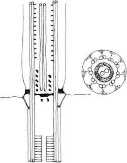
FIGURE 2.43 Type 4 transition zone of Chrysophyceae, Xantophyceae, and Eustigmatophyceae (Heterokontophyta)
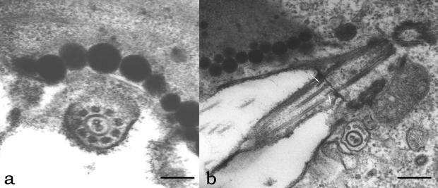
FIGURE 2.44 Transmission electron microscopy image of the transition zone of Ochromonas danica in transverse section (a) (Bar: 0.20 µm); longitudinal section of the short flagellum of Ochromonas (b); arrows point to the double rows of punctae representing the turn of the helix. (Bar: 0.40 µm.)
Type 5 (Figure 2.45) is characterized by the so-called “stellate pattern”; typically, it is divided into a longer distal and a shorter proximal part, separated by a basal plate. In longitudinal section, the structure resembles an H, with the cross-bar located a short distance above the cell surface (Figure 2.46). Transversely, the cross-bar may or may not extend to the peripheral doublets of the axoneme. Variations regard the length of the proximal part, the location of the plate, and the appearance of additional rings. This transition zone is typical of Chlorophyta.
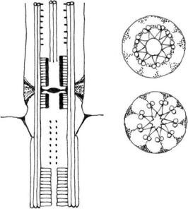
FIGURE 2.45 Type 5 transition zone (Chlorophyta).
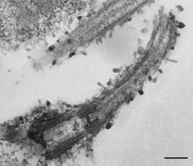
FIGURE 2.46 Transmission electron microscopy image of the transition zone of Dunaliella sp. in longitudinal section showing the “H” structure. (Bar: 0.40 µm.)


