Flagellar Shape and Surface Features

FIGURE 2.29 Transmission electron microscopy image of a Dunaliella sp. flagellum in transverse section, showing the homogeneous fuzzy coating of its membrane. (Bar: 0.10 µm)
FIGURE 2.29 Transmission electron microscopy image of a Dunaliella sp. flagellum in transverse section, showing the homogeneous fuzzy coating of its membrane. (Bar: 0.10 µm)
Flagella may bear a high variety of coverings and ornamentation, which often represent a taxonomic feature. The occurrence of flagellar scale follows that of cell body scales, because they are present only in eukaryotic algae, in the divisions of Heterokontophyta, Haptophyta, and Chlorophyta. As for the cell body scales, they have a silica-based composition in the Heterokonthophyta, a mixed structure of calcium carbonate and organic matter in the Haptophyta, and a completely organic nature in the Chlorophyta.
Members of the Chrysophyceae with flagellar scales (Heterokontophyta) fall into two groups: one possessing exactly the same type of scale on both flagellar and body surface, the other showing flagellar scale different in structure and arrangement from body scales. Example of the first group is Sphaleromantis sp., whose flagella and cell body are closely packed with scales of very peculiar appearance, resembling the branched structure of a tree. Examples of the second group are Mallomonas sp. and Synura sp.; in both genera, flagellar scales are not arranged in a regular pattern, are very small (under 300 nm) and possess different morphological types, the most characteristic being the annular type. As the body scales, flagellar scales are produced in deposition vesicles, extruded from the cell and brought into correct position in relation to the other scales and the cell surface.
Flagellar scales are known from almost all the genera of the class Prasinophyceae (Chlorophyta). These algae possess non-mineralized organic scales on their cell body and flagella, the same type of scale being rarely present on both surfaces. On the flagella, the scales are precisely arranged in parallel longitudinal rows, sometimes in one layer, two layers, or even three layers on top of each other. Each layer usually contains only one type of scales. The four flagella of Tetraselmis sp. are covered by different types of scales: pentagonal scales attached to the flagellar membrane (Figure 2.30), rod-shaped scales covering the pentagonal scales, and hair scales organized in two rows on opposite sides of the flagellum. A fourth type termed “knotted scales” is present only in some strains, but their precise arrangement is not known. In Nephroselmis spinosa the flagellar surface is coated by two different types of scales arranged in two distinct layers. Scales of the inner layer, deposited directly on the membrane, are small and square, 40 nm across (Figure 2.31); scales of the outer layer are rod-shaped, 30–40 nm long, and are deposited atop the inner scales. As in Tetraselmis, hair scales of at least two different types are also present covering the flagella.
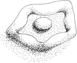
FIGURE 2.30 Pentagonal scale of the flagellar membrane of Tetraselmis sp.
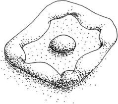
FIGURE 2.31 Square scale of the flagellar membrane of Nephroselmis spinosa.
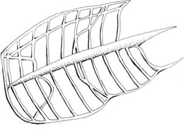
FIGURE 2.32 Limuloid scale of the flagellar membrane of Pyramimonas sp.
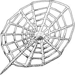
FIGURE 2.33 Spider web scale of the flagellar membrane of Mamiella gilva.
The scales are synthesized within the Golgi vesicles. The vesicles then migrate to the base of the flagella and from here are extruded and arranged on the flagella.
Support our developers


