The Nervous System: the Encephalon
In all vertebrated animals except Amphioxus, the brain exhibits that separation into a, fore-brain, mid-brain, and. hind-brain, which results from its embryonic division, by two constrictions, into the three thin-walled vesicles-the anterior, middle, and posterior cerebral vesicles - already mentioned. The cavities of these vesicles - the primitive ventricles of the brain - freely communicate at first, but become gradually diminished by the thickening of their sides and floors. The cavity of the anterior vesicle is, in the adult human brain, represented by the so-called third ventride; that of the middle vesicle, by the iter a. tertio ad quartum ventriculum ; that of the posterior vesicle, by the fourth verticle.The floor and sides of the posterior vesicle, in fact, thicken and become the medulla oblongata; together with the pons varolii, in those animals which possess the latter structure.
The posterior part of the roof is not converted into nervoua matter, but remains thin and attenuated ; the ependyma, or lining of the cerebral cavity, and the arachnoid, or serous membrane which covers the brain externally, coming nearly into contact, and forming, to all appearance, a single thin membrane, which tears with great readiness, and lays open the cavity of the fourth ventricle. Anteriorly, on the other hand, the roof becomes converted into nervous matter, and may enlarge into a complex mass, which overhangs the posterior division, and is called the cerebellum. The pons varolii, when it exists, is the expression of commissural fibres, which are developed in the sides and floor of the anterior part of the posterior cerebral vesicle, and connect one half of the cerebellum with the other.
Thus, the hind-brain differs from the posterior cerebral vesicle in being differentiated into the medulla oblongata (or myelencephalon) behind, and the cerebellum with the pons varolii (which together constitute the metencephalon) in front.
The floor of the middle cerebral vesicle thickens and becomes converted into two great bundles of longitudinal fibres, the crura cerebri. Its roof, divided into two, or four, convexities by a single longitudinal, or a crucial, depression, is converted into the " optic lobes," corpora bigemina or quadrigemina. And these parts, the optic lobes, the crura cerebri, and the interposed cavity, which either retains the form of a ventricle, or is reduced to a mere canal (the iter a tertio ad quartum ventriculum), are the components of the mid-brain or mesencephalon.
The anterior cerebral vesicle undergoes much greater changes than either of the foregoing; for, in the first place, it throws out from its anterior lateral parietes two hollow prolongations, the hemispheres (or prosencephala). and each of these again protrudes from its anterior end a smaller hollow process, the olfactory lobe (or rhinencephalon). By the development of these processes the anterior vesicle becomes divided into five parts
- one median and posterior, and four anterior and paired.
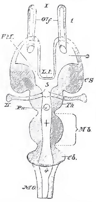 |
| Fig. 19. - Diagrammatic borizontal section of a Vertebrate braio.
The following letter
serve for both this figure and Fig. 20: Mb Mid-brain. What lies in front of this is the
fore-brain, and
what lies
behind, the hind-brain. Z. t. the lamina terminalis; Olf. the
olfactory lobes; IImp, the hemispheres; Th. E, the thalamencephalon; Pn, the pineal gland Py. the pituitary body; FM, the foramen of Monro: CS. the corpus striatum; Th, the optic thalamus; CQ the corpora quadrigemina; CC. the crura cerebri; Cb, the cerebellum ; PV, the pons varolii; MO, the medulla oblongata; I, olfactorii; II,optic; III, point of exit from the brain of the motores oculorum; IV of the pathetic; VI. of the abducentes; V - XII, origin of the other cerebral nerves. 1, olfactory ventricle 2, lateral ventricle; 3, third ventricle; 4, fourth ventricle; +, iter a tertio ad quartum centriculum. |
The front wall of the vesicle, in part, becomes the so-called lamina terminalis, which is the delicate anterior boundary of the third ventricle. In certain directions, however, it thickens and gives rise to three sets of fibres, one transverse and two vertical - the former lying in front of the latter. The transverse fibres pass on either side into the corpora striata, and constitute the anterior commissure which connects those bodies. The vertical fibres are the anterior pillars of the fornix, and they pass below into the floor of the third ventricle, and into the corpora mammillaria, when those structures are developed.
The outer and under wall of each cerebral hemisphere thickens and becomes the corpus striatum, a ganglionic structure which, from its origin, necessarily abuts against the outer and interior part of the optic thalamus. The line of demarcation between the two corresponds with the lower lip (taenia semicircularis) of the aperture of communication (called the foramen of Munro) between the third ventricle and the cavity of the cerebral hemisphere, which is now termed the lateral ventricle. In the higher Vertebrata, the upper lip of the foramen of Munro thickens, and becomes converted into a bundle of longitudinal fibres, which is continuous, anteriorly, with the anterior pillars ot the fornix before mentioned. Posteriorly these longitudinal fibres are continued backward and downward along the inner wall of the cerebral hemisphere, following the junction of the corpora striata and optic thalami, and pass into a thickening of the wall of the hemisphere, which projects into the lateral ventricle, and is called the hippocampus major. Thus a longitudinal commissural band of nervous - fibres, extending from the floor of the third ventricle to that of the lateral ventricle, and arching over the foramen of Munro, is produced. The fibres of opposite sides unite over the roof of the third ventricle, and constitute what is called the body of the fornix. Behind this union the bands receive the name of the posterior pillars of the fornix.
The optic thalami may be connected by a gray soft commissure; and a posterior commissure, consisting of transverse nerve - fibres, is generally developed between the posterior ends of the two thalami.
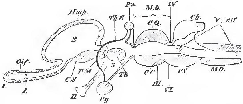 |
| Fig. 20. - A longitudinal and vertical section of a Vertebrate brain. - The letters as befora The lamina terrminalis is represented by the strong black line between FM and 3. |
When the corpus callosum is largely developed, its anterior part crosses the interspace between the hemispheres considerably above the level of the fornix; so that between the fornix and it, a certain portion of the inner wall of each hemisphere, with the intervening space, is intercepted. The portion of the two inner walls and their interspace, thus isolated from the rest, constitutes the septum lucidum, with its contained fifth ventricle.
Support our developers


