The Osseous Facial Apparatus
The bones of the face, which constitute the inferior arches of the skull, appear within the various processes and visceral arches which have been enumerated. Thus, the premaxillae are two bones developed in the oral part of the naso-frontal process, one on each side of the middle line, between the external nasal apertures, or anterior nares, and the anterior boundary of the mouth.Ossification occurs in the palato-pterygoid cartilage at two chief points, one in front and one behind. The anterior gives rise to the palatine bone, the posterior to the pterygoid. Outside these, several membrane bones may make their appearance in the same process. The chief of these is the maxilla, which commonly unites, in front, with the premaxilla. Behind the maxilla there may be a second, the jugal; and occasionally behind this lies a third, the quadrato-jugal.
Between the maxilla, the prefrontal and the premaxilla, another membrane bone, called lachrymal, from its ordinary relation to the lachrymal canal, is very generally developed; and one or more supro-orbital and post-orbital ossifications may be connected with the bony boundaries of the orbit.
When these and the postfrontal membrane bone are simultaneously developed, they form two series of bony splints attached to the lateral wall of the skull, one set above and one below the orbit, which converge to the lachrymal. The upper series (lachrymal, supra-orbital, post-frontal, squamosal), terminates posteriorly over the proximal end of the quadrate bone, or mandibular suspensorium. The lower series (lachrymal, maxillary, jugal, quadrato-jugal) ends over the distal end of that bone, with which the quadrato-jugal is connected. The two series are connected behind the orbit by the postorbital (when it exists), but more commonly by the union of the jugal with the post-frontal and squamosal. The Ichthyosauria, Chelonia, Crocodilia, and some Lacertilia, exhibit this double series of bones most completely.
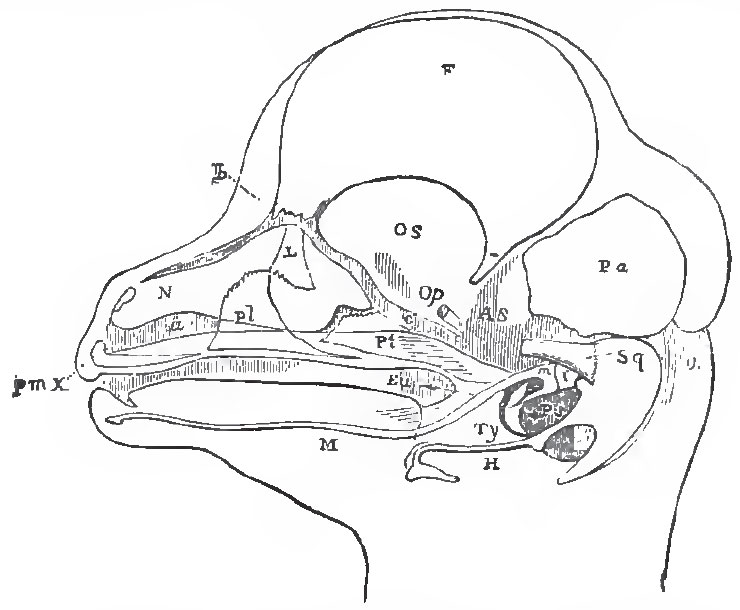 |
| Fig. 9. - The head of a fretal Lamb dissected so as to show Meckel's cartilage. M; thamalleus, m; the incus, i; the tympanic, Ty; the liyoid, II,' the squamosal, Sq; pterygold, Pt; palatine, pt, lachrymal, L; premaxilla, pme; nasal sac, N; Eustachin tube, Eu. |
Each nasal passage, at first very short, passes between the premaxilla below, the ethmoid and vomer on the inner side, the prefrontal above and externally, and the palatine behind, to open into the forepart of the mouth. And, before the cleft between the outer posterior angle of the naso-frontal process and the maxillary process is closed, this passage communicates laterally, with the exterior, and, posteriorly, with the cavity of the orbit. When the maxillary and the naso-frontal processes unite, the direct external communication ceases; but the orbito-nasal passage, or lachrymal canal, as it is called, in consequence of its function of conveying away the secretion of the lachrymal gland, may persist, and the lachrymal bone may be developed in especial relation with it.
In the higher Vertebrata, the nasal passages no longer communicate with the forepart of the cavity of the mouth; for the maxillaries and palatines, regularly, and the pterygoid bones, occasionally, send processes downward and inward, which meet in the middle line, and shut off from the mouth a canal which receives the nasal passages in front, while it opens, behind, into the pharynx, by what are now the posterior nares.
Two ossifications commonly appear near the proximal end of Meckel's cartilage, and become bones movably articulated together. The proximal of these is the quadrate bone found in most vertebrates, the malleus of mammals; the distal is the os articulare of the lower jaw in most vertebrates, but does not seem to be represented in mammals. The remainder of Meckel's cartilage usually persists for a longer or shorter time, but does not ossify. It becomes surrounded by bone, arising from one or several centres, in the adjacent membrane, and the ramus of the mandible thus formed articulates with the squamosal bone in mammals, but in other Vertebrata is immovably united with the os articulare. Hence the complete ramus of the mandible articulates directly with the skull in mammals, but only indirectly, or through the intermediation of the quadrate, in other Vertebrata.
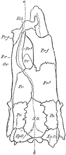 |
| Fig. 10 - The Skull of a plaice (Plastessa colguris) viewed from above. The logical median line; Or, Or, the position of the two eyes in theirs orbits; Eth, ethmoid; prf, prefontal: Fr. left frontal; Frl. right frontal; Pa, parietal; SO, supra-occipital; Ep.O, epiotic. |
The ossification of the hyoidean apparatus varies immensely in detail, but usually gives rise to bony lateral arches, and a median portion, bearing much the same relation to them as the sternum has to the ribs. When the lateral arches are complete, they are connected directly with the periotic capsule.
The proximal end of the hyoidean arch is often united, more or less closely, with the outer extremity of the bone called columella auris, or stapes, the inner end of which, in the higner Vertebrata, is attached to the membrane of the fenestra ovalis.
In ordinary fishes, a fold of the integument extends backward from the second visceral arch over the persistent branehial clefts; within this is developed a series of raylike membrane bones, termed opercular and branchiostegal, which be come closely connected with the hyoidean arch. A corresponding process of the skin is developed in the Batraohian Tadpole, and grows backward over the branchiae. Its posterior edge, at first free, eventually unites with the integument of the body, behind the branchial clefts, the union being completed much earlier on the right side than on the left.
In most mammals a similar fold of integument gives rise to the pinna, or external ear.
The branchial skeleton bears the same relation to the posterior visceral arches that the hyoidean does to the second. When fully developed, it exhibits ossified lateral arches, connected by median pieces, and frequently, provided with radiating appendages which give support to the branchial mucous membrane. It is only found in those Vertebrata which breathe by gills - the classes Pisces and Amphibia. In the higher Vertebrata, the posterior of the two pairs of cornua, with which the hyoidean apparatus is generally provided, are the only remains of the branchial skeleton.
The skull and face are usually symmetrical in reference to a median vertical plane. But, in some Cetacea, the bones about the region of the nose are unequally developed, and the skull becomes asymmetrical. In the Flatfishes (Pleuronectids), the skull becomes so completely distorted, that the two eyes lie on one side of the body, which is, in some caess, the left, and in other, the right side. In certain of thede fishes, the rest of the skull and facial bones, the spine, and even the limbs, partake in this asymmetry. The base of the skull and its occipital region are comparatively little affected; but, in the interorbital region, the frontal bones and the subjacent cartilaginous, or membranous, side-walls of the cranium are thrown over to one side; and, frequently, undergo a flexure, so that they become convex toward that side, and concave in the opposite direction. The prefontal bone of the side from which the skull is twisted, sends back a great process above the eye of that side, which unites with the frontal bone, and thus encloses this eye in a complete bony orbit. It is along this fronto-prefrontal bridge that the dorsal fin-rays are continued forward, just as if this bridge represented the morphological middle of the skull. (Fig. 10.)
The embryonic Pleuroneatidae have the eyes in their normal places, upon opposite sides of tlie head ; and the cranial distortion commences only after the fish are hatched.
Support our developers


