The Pectoral and Pelvic Arches
The proximal skeletal elements of each pair of limbs (humeri or femora) are supported by a primitively cartilaginous, pectoral, or pelvic girdle, which lies external to the costal elements of the vertebral skeleton. This girdle may consist of a simple cartilaginous arc (as in the Sharks and Rays), or it may be complicated by subdivisions and additions.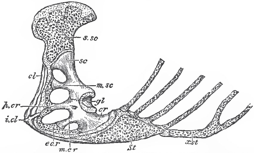 |
| Fig. 12. - Side-view of the pectoral arch and sternum of a Lizard (Iguana tuberculata).- Sc, scapula; 8.8C, supra-scapula; cr, coracoid; gl, glenoidal cavity; St, sternum; a.st, xiphisternum; m.sc, mesoscapula; p.cr, precoracoid; m.cr, mesocoracoid; e.cr, epicoracoid; cl, clavicle; i.cl, interclavicle. |
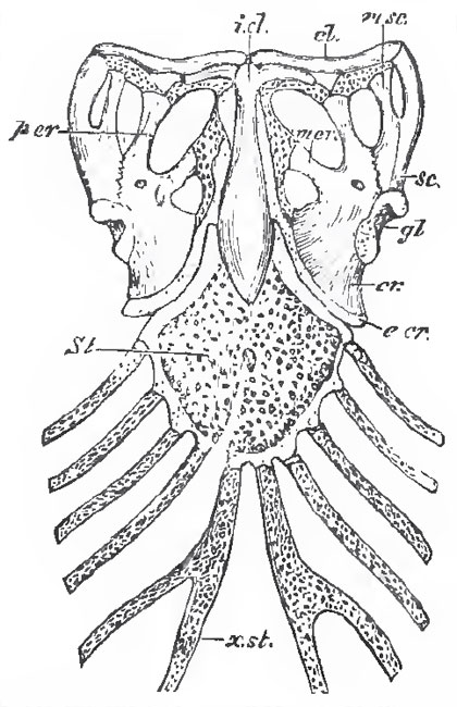 |
| Fig. 13. - Ventral view of the sternum and pectoral arches of Iguana tuberculata. The letters as in Fig. 12. |
In the great majority of the Vertebrata above fishes, the coracoids are large, and articulate with the antero-external margins of the primitively cartilaginous sternum, or breastbone. But, in most mammals, they do not reach the sternum, and, becoming anohylosed with the scapula, they appear, in adult life, as mere processes of that bone.
Numerous Vertebrates possess a clavicula, or collar-bone, which is connected with the pre-axial margin of the scapula and coracoid; but takes no part in the formation of the glenoid cavity, and is usually, if not always, a membrane bone. In many Vertebrata, the inner ends of the clavicles are connected with and supported by, a median membrane bone which is closely connected with a ventral face of the sternum. This is the interclavicula, frequently called episternum.
The pelvic, like the pectoral, arch at first consists of a simple continuous cartilage on each side, which, in Vertebrata higher than fishes, is divided by the acetabulum, or articular cavity for the reception of the head of the femur, into a dorsal and a ventral moiety.
Three separate ossifications usually take place in this cartilage-one in the dorsal, and two in the ventral, moiety. Hence, the pelvic arch eventually consists of a dorsal portion, called the ilium, and of two ventral elements, The pubis anteriorly, and the- ischium posteriorly. All these generally enter into the composition of the acetabulum.
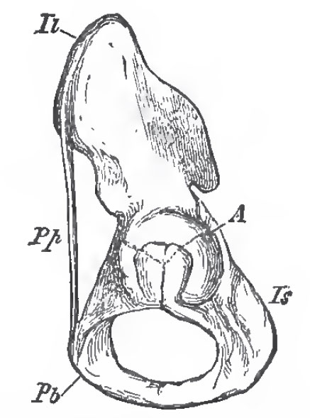 |
| Fig. 14 - Side-view of the left Os Innominatum of Man; Il, ilium; Ia, ischium; Pp, Pubic A, acetabulum; Pp, Poupart's ligament. |
The ischium corresponds very nearly with the coracoid in the pectoral arch; the pubis with the precoracoid, and more or less of the epicoracoid.
The pelvis possesses no osseous element corresponding with the clavicle, but a strong ligament, the so-called Poupartes ligament, stretches from the ilium to the pubis in many Vertebrata and takes its place. (Fig. 14, Pp.)
On the other hand, the marsupial bones of certain mammals, which are ossifications of the tendons of the external oblique muscles, seem to be unrepresented in the pectoral arch; while there appears to be nothing clearly corresponding with a sternum in the pelvic arch, though the precloacal cartilage, or ossicle, of Lizards has much the same relation to the isohia as the sternum has to the coracoids.
Very generally, though not universally, the ilia are closely articulated with the modified ribs of the sacrum. The pubes and ischia of opposite sides usually meet in a median ventral symphysis; but in all birds, except the Ostrich, this union does not take place.
Support our developers


