Human chromosomes

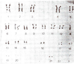
Fig. 24.2. G-banding karyotypes of human chromosomes showing linear differentiation and intercalary bands.

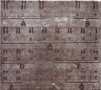
Fig. 24.3. C-banding karyotypes of human chromosomes showing intense bands at the centromeres.
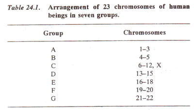
In several topics of genetics on Biocyclopedia.com, examples from human beings were utilized wherever available. However, the technique, developed by J.H. Tjio and A. Levan in 1956, enabled the determination of correct chromosome number (2n = 46) in human beings for the first time. Later, on the basis of morphology, twenty three chromosomes in human beings were arranged in seven groups (Table 24.1). Chromosomes of a human male at metaphase are shown in Figure 24.1. From such photographs, karyotypes are prepared according to the arrangement shown in Table 24.1.
During the last 20-25 years, human chromosomes have been studied using the new techniques of G-banding, C-banding and several other banding techniques. These techniques use specific dyes, which are sometimes fluorescent and which reveal ultrastructural differences between different human chromosomes. When subjected to certain treatments and then stained with chemical dye Giemsa, chromosomes generate a series of bands (Fig. 24.2). These banding techniques help in identification of each chromosome and any structural changes which might have occurred. The G-bands reveal the intercalary bands and C-bands reveal only the centromeric regions consisting mainly of constitutive rather than the facultative heterochromatin as shown in Figure 24.3 (see Physical Basis of Heredity 1. The Nucleus and the Chromosome.


