Genetics / Human Genetics
Sickle-cell anaemia
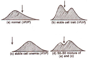
Fig. 24.4. Electrophoretic mobilities in normal and abnormal haemoglobins.
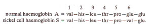
Fig. 24.5. Segment of β chains of normal and abnormal haemoglobin showing amino acid replacement.
Sickle cell anaemia is a blood disease where the red blood cells become sickle shaped as compared with round shape in normal individual. This results into various abnormalities and may ultimately result into death. This disease is caused by a single gene, which in heterozygous condition causes moderate sickling (sickle cell trait) and in homozygous condition causes severe effect (sickle cell anaemia). It was also found that the haemoglobins of normal individual and a patient have different mobilities in an electrophoretic field (Fig. 24.4). Haemoglobin of sickle cell anaemia moves in a direction opposite to that of normal haemoglobin. Such a difference was later discovered to be due to the replacement of a single amino acid in βchain of haemoglobin (Fig. 24.5).
Fig. 24.4. Electrophoretic mobilities in normal and abnormal haemoglobins.
Fig. 24.5. Segment of β chains of normal and abnormal haemoglobin showing amino acid replacement.
Support our developers

More in this section


