Prokaryotic Nucleoids
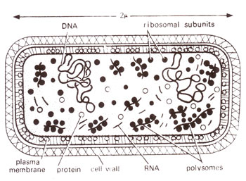
Fig. 6.19. A bacterial cell showing details of internal structure (redrawn from De Robertis et al., Cell Biology).
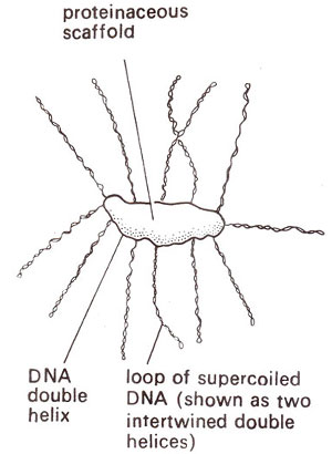
Fig. 6.20. Diagrammatic representation of the organization of DNA and proteins in bacterial chromosome.
These bodies can be clearly observed with the help of electron microscope. It has also been demonstrated that these bodies consist of a network of fine threads (Fig. 6.19). Recent work by a number of workers demonstrated that the network of threads consists of a single chromosome in the form of a ring. The exact three dimensional arrangement by which 1100μ -1400μ long DNA chain, which forms 80% of the chromosome by mass (the remaining 20% being protein + RNA), is packed in a lμ long nucleoid, could also be established now.
Fig. 6.19. A bacterial cell showing details of internal structure (redrawn from De Robertis et al., Cell Biology).
Fig. 6.20. Diagrammatic representation of the organization of DNA and proteins in bacterial chromosome.
Support our developers


