Meiosis: Reduction Division of Gametes
Meiosis: Reduction
Division of Gametes
Every body cell contains two chromosomes
bearing genes for the same set
of characteristics, and the two members
of each pair usually, but not
always, have the same size and shape.
The members of such a pair are called homologous chromosomes; one of
each pair comes from the mother and
the other from the father. Meiosis consists
of two nuclear divisions in which
the chromosomes divide only once
(Figure 5-2). The result is that mature
gametes have only one member of
each homologous chromosome pair,
or a haploid (n) number of chromosomes.
When the gametes unite in any
fertilization, a zygote is formed. In
humans the zygotes and all body cells
normally have the diploid number
(2n), or 46 chromosomes; the gametes
have the haploid number (n), or 23,
and meiosis reduces the number of
chromosomes from diploid to haploid.
Alleles are alternative forms of the same gene that have arisen by mutation of the DNA sequence. Like a baseball team with several pitchers, only one of whom can occupy the pitcher’s mound at a time, only one allele can occupy a chromosomal locus. Alternative alleles for the locus may be on homologous chromosomes of a single individual, making that individual heterozygous for the gene in question. Numerous allelic forms of a gene may be found among different individuals in the population of the species.
During an individual’s growth, all the chromosomes of the mitotically dividing cells contain the double set of chromosomes. In the reproductive organs, the gametes (germ cells) are formed after meiosis, which separates the homologous pairs of chromosomes. If it were not for this reductional division, the union of ovum (egg) and sperm would produce an individual with twice as many chromosomes as the parents. Continuation of this process in just a few generations could yield astronomical numbers of chromosomes per cell.
Most of the unique features of meiosis occur during the prophase of the first meiotic division (Figure 5-2). The two members of each pair of homologous chromosomes come into side-by-side contact (synapsis) to form a bivalent. Each chromosome of the bivalent has already replicated to form two chromatids, each of which will become a new chromosome. The two chromatids are joined at one point, the centromere, so that each bivalent is composed of two pairs of chromatids (each pair is a dyad), or four future chromosomes, and is thus called a tetrad. The position or location of any gene on a chromosome is the gene locus (pl., loci), and in synapsis all gene loci on a chromatid normally lie exactly opposite the corresponding loci on the sister chromatid. Toward the end of the prophase, the chromosomes shorten and thicken and are ready to enter into the first meiotic division. In contrast to mitosis, the centromeres holding the chromatids together do not divide at the beginning of anaphase. As a result, each of the dyads is pulled toward each pole by the microtubules of the division spindle. Therefore at the end of the first meiotic division, the daughter cells contain one of each of the homologous chromosomes, so the total chromosome number has been reduced to haploid. However, because the chromatids are still joined by their centromeres, each cell contains 2n amount of DNA.
The second meiotic division more closely resembles the events in mitosis. The dyads are split at the beginning of anaphase by division of the centromeres, and single-stranded chromosomes move toward each pole. Thus by the end of the second meiotic division, the cells have the haploid number of chromosomes and n amount of DNA. Each chromatid of the original tetrad exists in a separate nucleus. Four products are formed, each containing one complete haploid set of chromosomes and only one allele of each gene. Only one of the four products in female gametogenesis will become a functional gamete.
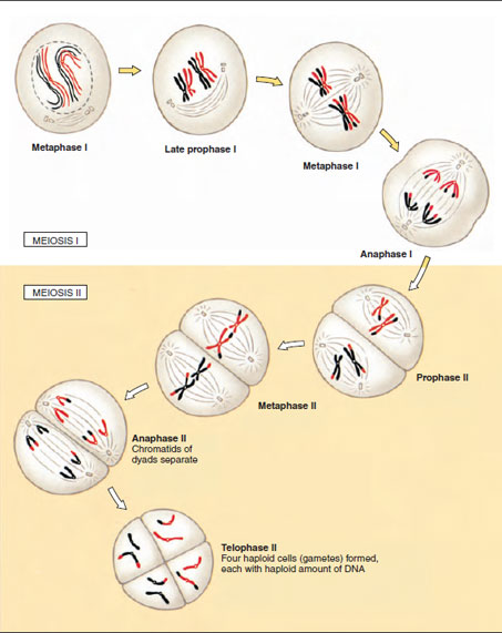 |
| Figure 5-2 Meiosis in a sex cell with two pairs of chromosomes. |
Alleles are alternative forms of the same gene that have arisen by mutation of the DNA sequence. Like a baseball team with several pitchers, only one of whom can occupy the pitcher’s mound at a time, only one allele can occupy a chromosomal locus. Alternative alleles for the locus may be on homologous chromosomes of a single individual, making that individual heterozygous for the gene in question. Numerous allelic forms of a gene may be found among different individuals in the population of the species.
During an individual’s growth, all the chromosomes of the mitotically dividing cells contain the double set of chromosomes. In the reproductive organs, the gametes (germ cells) are formed after meiosis, which separates the homologous pairs of chromosomes. If it were not for this reductional division, the union of ovum (egg) and sperm would produce an individual with twice as many chromosomes as the parents. Continuation of this process in just a few generations could yield astronomical numbers of chromosomes per cell.
Most of the unique features of meiosis occur during the prophase of the first meiotic division (Figure 5-2). The two members of each pair of homologous chromosomes come into side-by-side contact (synapsis) to form a bivalent. Each chromosome of the bivalent has already replicated to form two chromatids, each of which will become a new chromosome. The two chromatids are joined at one point, the centromere, so that each bivalent is composed of two pairs of chromatids (each pair is a dyad), or four future chromosomes, and is thus called a tetrad. The position or location of any gene on a chromosome is the gene locus (pl., loci), and in synapsis all gene loci on a chromatid normally lie exactly opposite the corresponding loci on the sister chromatid. Toward the end of the prophase, the chromosomes shorten and thicken and are ready to enter into the first meiotic division. In contrast to mitosis, the centromeres holding the chromatids together do not divide at the beginning of anaphase. As a result, each of the dyads is pulled toward each pole by the microtubules of the division spindle. Therefore at the end of the first meiotic division, the daughter cells contain one of each of the homologous chromosomes, so the total chromosome number has been reduced to haploid. However, because the chromatids are still joined by their centromeres, each cell contains 2n amount of DNA.
The second meiotic division more closely resembles the events in mitosis. The dyads are split at the beginning of anaphase by division of the centromeres, and single-stranded chromosomes move toward each pole. Thus by the end of the second meiotic division, the cells have the haploid number of chromosomes and n amount of DNA. Each chromatid of the original tetrad exists in a separate nucleus. Four products are formed, each containing one complete haploid set of chromosomes and only one allele of each gene. Only one of the four products in female gametogenesis will become a functional gamete.
Support our developers


