Peripheral Nervous System
Peripheral Nervous System
The peripheral nervous system includes all nervous tissue outside the central nervous system. It consists of two functional divisions: sensory or afferent division, which brings sensory information to the central nervous system, and motor or efferent division, which conveys motor commands to muscles and glands. The efferent division consists of two components: (1) somatic nervous system, which innervates skeletal muscle, and (2) autonomic nervous system, which innervates smooth muscle, cardiac muscle, and glands.
Autonomic Nervous System The autonomic system governs involuntary, internal functions of the body that do not ordinarily affect consciousness, such as movements of the alimentary canal and heart, contraction of the smooth muscle of blood vessels, urinary bladder, iris of the eye, and others, plus secretions of various glands.
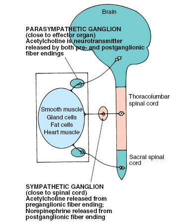
Autonomic nerves originate in the brain or spinal cord as do nerves of the somatic nervous system, but unlike the latter, autonomic fibers consist of not one but two motor neurons. They synapse once after leaving the cord and before arriving at the effector organ. These synapses are located outside the spinal cord in ganglia. Fibers passing from the cord to the ganglia are called preganglionic autonomic fibers; those passing from the ganglia to the effector organs are called postganglionic fibers. These relationships are illustrated in Figure 35-16.
Subdivisions of the autonomic system are the parasympathetic and sympathetic systems. Most organs in the body are innervated by both sympathetic and parasympathetic fibers, whose actions are antagonistic (Figure 35-17). If one fiber stimulates an activity, the other inhibits it. However, neither kind of nerve is exclusively excitatory or inhibitory. For example, parasympathetic fibers inhibit heartbeat but excite peristaltic movements of the intestine; sympathetic fibers increase heartbeat but inhibit intestinal peristaltic movement.
The parasympathetic system consists of motor neurons, some of which emerge from the brain stem by certain cranial nerves and others of which emerge from the sacral (pelvic) region of the spinal cord (Figures 35-16 and 35-17). In the sympathetic division nerve cell bodies of all the preganglionic fibers are located in the thoracic and upper lumbar areas of the spinal cord. Their fibers exit through the ventral roots of the spinal nerves, separate from these, and go to sympathetic ganglia (Figure 35-17), which are paired and form a chain on each side of the spinal column.
All preganglionic fibers, whether sympathetic or parasympathetic, release acetylcholine at the synapse with postganglionic cells. However, parasympathetic postganglionic fibers release acetylcholine at their endings, whereas sympathetic postganglionic fibers with few exceptions release norepinephrine (also called noradrenaline). This difference is another important characteristic distinguishing the two parts of the autonomic nervous system.
As a general rule the parasympathetic division is associated with nonstressful activities, such as resting, eating, digestion, and urination. The sympathetic division is active under conditions of physical or emotional stress. Under such conditions heart rate increases, blood vessels to the skeletal muscles dilate, blood vessels in the viscera constrict, activity of the intestinal tract decreases, and metabolic rate increases. The importance of these responses in emergency reactions (sometimes called the fright, fight or flight response) are described in the next section. It should be noted, however, that the sympathetic division is active also during resting conditions in maintaining normal blood pressure and body temperature.
 |
| Figure 35-16 General organization of the autonomic nervous system |
The peripheral nervous system includes all nervous tissue outside the central nervous system. It consists of two functional divisions: sensory or afferent division, which brings sensory information to the central nervous system, and motor or efferent division, which conveys motor commands to muscles and glands. The efferent division consists of two components: (1) somatic nervous system, which innervates skeletal muscle, and (2) autonomic nervous system, which innervates smooth muscle, cardiac muscle, and glands.
Autonomic Nervous System The autonomic system governs involuntary, internal functions of the body that do not ordinarily affect consciousness, such as movements of the alimentary canal and heart, contraction of the smooth muscle of blood vessels, urinary bladder, iris of the eye, and others, plus secretions of various glands.
Autonomic nerves originate in the brain or spinal cord as do nerves of the somatic nervous system, but unlike the latter, autonomic fibers consist of not one but two motor neurons. They synapse once after leaving the cord and before arriving at the effector organ. These synapses are located outside the spinal cord in ganglia. Fibers passing from the cord to the ganglia are called preganglionic autonomic fibers; those passing from the ganglia to the effector organs are called postganglionic fibers. These relationships are illustrated in Figure 35-16.
Subdivisions of the autonomic system are the parasympathetic and sympathetic systems. Most organs in the body are innervated by both sympathetic and parasympathetic fibers, whose actions are antagonistic (Figure 35-17). If one fiber stimulates an activity, the other inhibits it. However, neither kind of nerve is exclusively excitatory or inhibitory. For example, parasympathetic fibers inhibit heartbeat but excite peristaltic movements of the intestine; sympathetic fibers increase heartbeat but inhibit intestinal peristaltic movement.
The parasympathetic system consists of motor neurons, some of which emerge from the brain stem by certain cranial nerves and others of which emerge from the sacral (pelvic) region of the spinal cord (Figures 35-16 and 35-17). In the sympathetic division nerve cell bodies of all the preganglionic fibers are located in the thoracic and upper lumbar areas of the spinal cord. Their fibers exit through the ventral roots of the spinal nerves, separate from these, and go to sympathetic ganglia (Figure 35-17), which are paired and form a chain on each side of the spinal column.
All preganglionic fibers, whether sympathetic or parasympathetic, release acetylcholine at the synapse with postganglionic cells. However, parasympathetic postganglionic fibers release acetylcholine at their endings, whereas sympathetic postganglionic fibers with few exceptions release norepinephrine (also called noradrenaline). This difference is another important characteristic distinguishing the two parts of the autonomic nervous system.
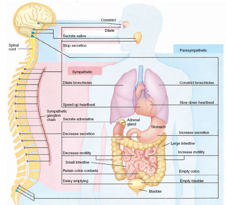 |
| Figure 35-17 Autonomic nervous system in humans. Outflow of autonomic nerves from the central nervous system is shown at left. Sympathetic (red) outflow is from the thoracic and lumbar areas of the spinal cord by way of a chain of sympathetic ganglia. Parasympathetic (blue) outflow is from the cranial and sacral regions of the central nervous system; parasympathetic ganglia (not shown) are located in or adjacent to the organs innervated. Most organs are innervated by fibers from both sympathetic and parasympathetic divisions. |
As a general rule the parasympathetic division is associated with nonstressful activities, such as resting, eating, digestion, and urination. The sympathetic division is active under conditions of physical or emotional stress. Under such conditions heart rate increases, blood vessels to the skeletal muscles dilate, blood vessels in the viscera constrict, activity of the intestinal tract decreases, and metabolic rate increases. The importance of these responses in emergency reactions (sometimes called the fright, fight or flight response) are described in the next section. It should be noted, however, that the sympathetic division is active also during resting conditions in maintaining normal blood pressure and body temperature.
Support our developers


