Cell Count by Hemocytometer or Measuring Volume
- Microscope
- Hemocytometer and coverslip
- Suspension of yeast
Procedure
- Make a serial dilution series of the yeast suspension, from 1/10 to 1/1000.
- Obtain a hemocytometer and place it on the desk before you. Place a clean coverslip over the center chamber.
- Starting with the 1/10 dilution, use a Pasteur pipette to transfer a small aliquot of the dilution to the hemocytometer. Place the tip of the pipette into the V-shaped groove of the hemocytometer and allow the cell suspension to flow into the chamber of the hemocytometer by capillary action until the chamber is filled. Do not overfill the chamber.

Figure 12 Hemocytometer.
- Add a similar sample of diluted yeast to the opposite side of the chamber and allow the cells to settle for about 1 minute before counting.
- Refer to the diagram of the hemocytometer grid in Figure 12 and note the following.
- The coverslip is 0.1 mm above the grid, and the lines etched on the grid are at preset dimensions.
- The 4 outer squares, marked 1-4, each cover a volume of 10–4 mL.
- The inner square, marked as 5, also covers a volume of 10–4 mL, but is further subdivided into 25 smaller squares. The volume over each of the 25 smaller squares is 4.0 × 10⁻⁶ mL.
- Each of the 25 smaller squares is further divided into 16 squares, which are the smallest gradations on the hemocytometer. The volume over these smallest squares is .25 × 10–6 mL.
- Given these volumes, the number of cells in a sample can be determined by counting the number of cells in one or more of the squares. Which square to use depends on the size of the object to be counted. Whole cells would use the larger squares, counted with 10X magnification. Isolated mitochondria would be counted in the smallest squares with at least 40X magnification.
- For the squares marked 1–4, the area of each is 1 mm2, and the volume is .1 mm3. Since .1 mm3 equals 10–4 mL, the number of cells/mL = average number of cells per 1 mm2 × 104 × any sample dilution.
- For the 25 smaller squares in the center of the grid marked 5, each small square is 0.2 × 0.2 mm2, and the volume is thus 0.004 mm3. For small cells, or organelles, the particles/mL equals the average number of particles per small square × 25 × 104 × any sample dilution.
- Grids 1–5 are all 1 mm2. Grids 1–4 are divided into 16 smaller squares (0.25 mm on each side), and grid 5 is divided into 25 smaller squares (0.2 mm on each side). Grid 5 is further subdivided into 16 of the smallest squares found on the hemocytometer.
- For the yeast suspension, count the number of cells in 5 of the intermediate, smaller squares of the hemocytometer. For statistical validity, the count should be between 10 and 100 cells per square. If the count is higher, clean out the hemocytometer and begin again with step 3, but use the next dilution in the series.
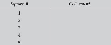
Record the dilution used, and the 5 separate counts. Average your counts, multiply by the dilution factor, and calculate the number of cells/mL in the yeast suspension. Record this information in the space provided.

Area of each square = __________ mm2 × 0.1 mm depth
= volume of each square.
Volume of each square = __________ mm3
Average number of cells per mm3 = __________
Number of cells per cm3 (1000 × above) = __________
Number of cells per cm3 is also number per mL.
Number of cells per mL __________ × dilution factor (200) = __________
cells per mL of whole blood.


