Medically Significant Gram–Positive Cocci (GPC)
- Perform, interpret, and define the relevance of the catalase test.
- Perform, interpret, and define the relevance of the coagulase test.
- Perform, interpret, and define the relevance of the test for hemolysis on BAP.
- Describe the Gram stain and arrangement of major GPC families.
- Determine the identification of an unknown GPC organism using the above tests.
- Relate the medical significance of each of the GPC covered in the lab.
Medically significant Gram-positive cocci are represented by 2 main families: Micrococcaceae (including the genera Staphylococcus and Micrococcus) and Streptococcaceae (including the genera Streptococcus and Enterococcus).
Micrococcaceae
Catalase +, Gram-positive cocci in clusters.
- Genus Micrococcus. These bacteria are rarely associated with disease and are common environmental contaminants. They Gram-stain as GPC in tetrads and produce yellow (Micrococcus luteus) or rose (M. roseus)—colored pigments on enriched media.
- Genus Staphylococci—Salt-tolerant GPC
- Staphylococcus aureus—These are pathogenic bacteria causing wound infections, abscesses, carbuncles, bacteremia, septicemia, and osteomyelitis. Associated with purulent discharges and capable of producing a wide range of exotoxins (including hemolysins and DNAse), characterized as highly invasive. Causes food poisoning by the production of a heat-resistant toxin. Important in nosocomial infections, especially MRSA (methicillin- or multiple-resistant Staphylococcus aureus). Nasal carriers are important.
- Staphylococcus epidermidis—Normal skin flora, nonhemolytic, coagulasenegative. Opportunistic pathogen isolated from catheters and IV lines and associated with transplant and immunosuppressed patients.
- Staphylococcus hemolyticus—Normal skin inhabitant, beta-hemolytic but coagulase negative. Opportunistic pathogen associated with UTIs.
Streptococcaceae
Gram-positive cocci in chain and pairs, easily decolorized. This family produces a large number of exotoxins including hemolysins, erythrogenic toxins, nephrotoxins, and cardiohepatic toxins. Pathogenesis depends on species, strain, portal of entry, and immune response. The Streptococcaceae are fastidious, requiring blood agar for growth and producing typical characteristics as the blood cells are destroyed (hemolysis) for nutrients.
- Streptococci. Chains and pairs, some encapsulated, bile-, esculin-, and saltnegative
- Strep. pyogenes—Group A strep, beta-hemolytic, causing strep throat, scarlet fever, peurperal fever (postnatal sepsis), skin infections such as impetigo, and pneumonia. Post-infection sequelae such as glomerulonephritis and rheumatic fever represent serious syndromes if infections are not treated immediately.
- Strep. agalactiae—Group B beta-hemolytic strep, causing neonatal meningitis thought to be associated with asymptomatic vaginal carriers. Recently reported in AIDS patients.
- Strep. pneumoniae—Alpha-hemolytic, mucoid, and lancet-shaped. Virulent strains are encapsulated. Causes pneumonia, ear, and eye infections.
- Alpha strep—Variety of nonpathogenic normal flora found on the skin and in the mouth. Occasionally associated with bacterial endocarditis. Many of these alpha strep are found on the respiratory tract as normal flora. Strep mutans is one of these strep associated with dental caries. Strep. sanguinis and Strep. parasanguinis are normal oral flora.
- Enterococci. Salt-tolerant bile esculin-positive strep. Normal flora of the GI tract. Opportunistic pathogens infecting decubiti (bedsores), causing UTIs and associated with IV contamination. The enterococci are medically significant due to growing antibiotic resistance, they are referred to as VRE (vancomycin-resistant enterococci). Many species make up this group of strep, including Enterococcus faecalis and Enterococcus faecium.
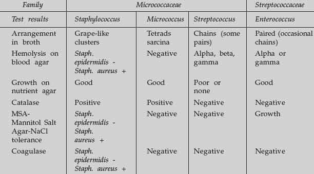
The following table represents a simple differentiation between these genera of bacteria.
Materials
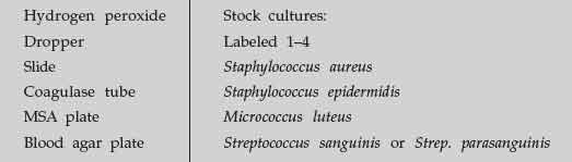 |
 |
Figure 52 MSA plate divided into quadrants. |
Procedure
- Gram-stain each of the stock cultures and record the results.
- Divide a blood agar plate into 4 sections and label them. Inoculate each section with a streak from each stock culture.
- Perform a catalase test by transferring a loopful of bacteria from each stock culture to a glass slide. Using a dropper apply a drop of hydrogen peroxide to each bacterial smear and record the results. Bubbling = positive.
- Divide an MSA plate into quadrants and inoculate it in the same fashion that the BAP was inoculated.
- Perform a coagulase test, using the premade tubes and inoculating a tube with each of the stock cultures. Cover the tube with parafilm and incubate for 24–48 hours.
- Incubate the BAP plate and MSA plate, and coagulase at 37°C. During the next lab period, read and record the results.
- Determine the identity of each stock culture.
Result
Observe your experimental result and interpret it.
Exercise
- Define the following terms and describe a positive test.
beta hemolysis -
alpha hemolysis -
gamma hemolysis - - Describe a positive coagulase test and identify the species that would produce a positive test.
- What advantage might coagulase provide for a pathogen as it invades the human body?
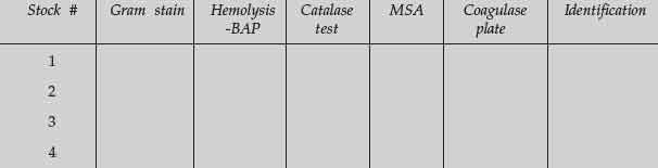
- Fill in the chart below with your results and determine the identity of each of yo ur stock cultures as a lab team.
Support our developers


