Nephridium
Nephridium
The most common type of invertebrate excretory organ is the nephridium, a tubular structure designed to maintain appropriate osmotic balance. One of the simplest arrangements is the flame cell system (or protonephridium) of acoelomates (flatworms) and some pseudocoelomates.
In planaria and other flatworms the protonephridial system takes the form of two highly branched duct systems distributed throughout the body (Figure 32-5). Fluid enters the system through specialized “flame cells,” moves slowly into and down the tubules, and is excreted through pores that open at intervals on the body surface. The rhythmical beat of the flagellar tuft, suggestive of a tiny flickering flame, creates a negative pressure that draws fluid into the tubular portion of the system. In the tubule, water and metabolites valuable to the body are recovered by reabsorption, leaving wastes behind to be expelled. Nitrogenous wastes (mainly ammonia) diffuse across the surface of the body.
The flame-cell system is extensively branched throughout a flatworm’s body because these acoelomate animals have no circulatory system to deliver wastes to a centralized excretory system (such as the kidneys of vertebrates and many invertebrates).
The protonephridium just described is a closed system. The tubules are closed on the inner end and urine is formed from a fluid that must first enter the tubules by being transported across flame cells. A more advanced type of nephridium is the open, or “true,” nephridium (metanephridium) that is found in several eucoelomate phyla such as annelids (Figure 32-6), molluscs, and several smaller phyla. A metanephridium is more advanced than a protonephridium in two important ways. First, the tubule is open at both ends, allowing fluid to be swept into the tubule through a ciliated funnellike opening, the nephrostome. Second, a metanephridium is surrounded by a network of blood vessels that assists in the reclamation of water and valuable materials such as salts, sugars, and amino acids from the tubular fluid.
Despite these differences, the basic process of urine formation is the same in protonephridia and metanephridia: fluid enters and flows continuously through a tubule where the fluid is selectively modified by (1) withdrawing valuable solutes from it and returning these to the body (reabsorption) and (2) adding waste solutes to it (secretion). The sequence ensures removal of wastes from the body without loss of materials valuable to the body. We will see that kidneys of vertebrates operate in basically the same way.
Arthropod Kidneys
The paired antennal glands of crustaceans, located in the ventral part of the head (Figure 32-7), are an advanced design of the basic nephridial organ. However, they lack open nephrostomes. Instead, hydrostatic pressure of the blood forms a proteinfree filtrate of the blood (ultrafiltrate) in the end sac. In the tubular portion of the gland, selective reabsorption of certain salts and active secretion of others modifies the filtrate. Thus crustaceans have excretory organs that are basically vertebrate-like in the functional sequence of urine formation.
Insects and spiders have a unique excretory system consisting of Malpighian tubules that operate in conjunction with specialized glands in the wall of the rectum (Figure 32-8). These thin, elastic, blind Malpighian tubules are closed and lack an arterial supply. Urine formation is initiated by active secretion of salts, largely potassium, into the tubules from the hemolymph (blood). This primary secretion of ions creates an osmotic drag that pulls water, solutes, and nitrogenous wastes, especially uric acid, into the tubule. Uric acid enters the upper end of the tubule as soluble potassium urate, which precipitates as insoluble uric acid in the proximal end of the tubule. Once the formative urine drains into the rectum, most of the water and potassium are reabsorbed by specialized rectal glands, leaving behind uric acid and other wastes that are expelled in the feces. The Malpighian tubule excretory system is ideally suited for life in dry environments and has contributed to the adaptive radiation of insects on land.
The most common type of invertebrate excretory organ is the nephridium, a tubular structure designed to maintain appropriate osmotic balance. One of the simplest arrangements is the flame cell system (or protonephridium) of acoelomates (flatworms) and some pseudocoelomates.
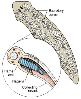 |
| Figure 32-5 Flame cell system of a flatworm. Body fluids collected by flame cells (protonephridia) are passed down a system of ducts to excretory pores on the body surface |
In planaria and other flatworms the protonephridial system takes the form of two highly branched duct systems distributed throughout the body (Figure 32-5). Fluid enters the system through specialized “flame cells,” moves slowly into and down the tubules, and is excreted through pores that open at intervals on the body surface. The rhythmical beat of the flagellar tuft, suggestive of a tiny flickering flame, creates a negative pressure that draws fluid into the tubular portion of the system. In the tubule, water and metabolites valuable to the body are recovered by reabsorption, leaving wastes behind to be expelled. Nitrogenous wastes (mainly ammonia) diffuse across the surface of the body.
The flame-cell system is extensively branched throughout a flatworm’s body because these acoelomate animals have no circulatory system to deliver wastes to a centralized excretory system (such as the kidneys of vertebrates and many invertebrates).
The protonephridium just described is a closed system. The tubules are closed on the inner end and urine is formed from a fluid that must first enter the tubules by being transported across flame cells. A more advanced type of nephridium is the open, or “true,” nephridium (metanephridium) that is found in several eucoelomate phyla such as annelids (Figure 32-6), molluscs, and several smaller phyla. A metanephridium is more advanced than a protonephridium in two important ways. First, the tubule is open at both ends, allowing fluid to be swept into the tubule through a ciliated funnellike opening, the nephrostome. Second, a metanephridium is surrounded by a network of blood vessels that assists in the reclamation of water and valuable materials such as salts, sugars, and amino acids from the tubular fluid.
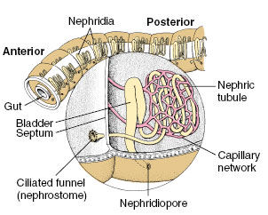 |
| Figure 32-6 Excretory system of an earthworm. Each segment has a pair of large nephridia suspended in a fluid-filled coelom. Each nephridium occupies two segments because the ciliated funnel (nephrostome) drains the segment anterior to the segment containing the rest of the nephridium. |
Despite these differences, the basic process of urine formation is the same in protonephridia and metanephridia: fluid enters and flows continuously through a tubule where the fluid is selectively modified by (1) withdrawing valuable solutes from it and returning these to the body (reabsorption) and (2) adding waste solutes to it (secretion). The sequence ensures removal of wastes from the body without loss of materials valuable to the body. We will see that kidneys of vertebrates operate in basically the same way.
Arthropod Kidneys
The paired antennal glands of crustaceans, located in the ventral part of the head (Figure 32-7), are an advanced design of the basic nephridial organ. However, they lack open nephrostomes. Instead, hydrostatic pressure of the blood forms a proteinfree filtrate of the blood (ultrafiltrate) in the end sac. In the tubular portion of the gland, selective reabsorption of certain salts and active secretion of others modifies the filtrate. Thus crustaceans have excretory organs that are basically vertebrate-like in the functional sequence of urine formation.
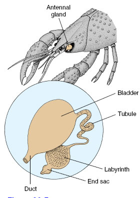 |
| Figure 32-7 Antennal glands of a crayfish. These are filtration kidneys in which a filtrate of the blood is formed in the end sac. The filtrate is converted into urine as it passes down the tubule toward the bladder |
Insects and spiders have a unique excretory system consisting of Malpighian tubules that operate in conjunction with specialized glands in the wall of the rectum (Figure 32-8). These thin, elastic, blind Malpighian tubules are closed and lack an arterial supply. Urine formation is initiated by active secretion of salts, largely potassium, into the tubules from the hemolymph (blood). This primary secretion of ions creates an osmotic drag that pulls water, solutes, and nitrogenous wastes, especially uric acid, into the tubule. Uric acid enters the upper end of the tubule as soluble potassium urate, which precipitates as insoluble uric acid in the proximal end of the tubule. Once the formative urine drains into the rectum, most of the water and potassium are reabsorbed by specialized rectal glands, leaving behind uric acid and other wastes that are expelled in the feces. The Malpighian tubule excretory system is ideally suited for life in dry environments and has contributed to the adaptive radiation of insects on land.
Support our developers


