Structure of Chromosomes
Structure of Chromosomes
As mentioned earlier, DNA in eukaryotic
cells occurs in chromatin, a complex
of DNA with histone and nonhistone
protein. Chromatin is organized
into a number of discrete bodies called
chromosomes (color bodies), so
named because they stain deeply with
certain biological dyes. In cells that are
not dividing, chromatin is loosely organized
and dispersed, so that individual
chromosomes cannot be distinguished
(Principles of Genetics:A Review). Before division the
chromatin condenses, and chromosomes
can be recognized and their
individual morphological characteristics
determined. They are of varied
lengths and shapes, some bent and
some rodlike. Their number is constant
for the species, and every body cell
(but not the germ cells) has the same
number of chromosomes regardless of
the cell’s function. A human, for example,
has 46 chromosomes in each
somatic cell.
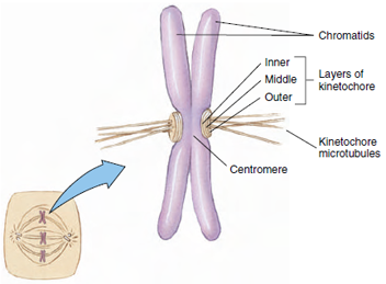
During mitosis (nuclear division) chromosomes shorten and become increasingly condensed and distinct, and each assumes a shape partly characterized by the position of a constriction, the centromere (Figure 3-21). The centromere is the location of the kinetochore, a disc of proteins specialized to bind with microtubules of the spindle fibers during mitosis.
The problem of packaging the cell’s DNA so that the genetic instructions are accessible during the transcription process is formidable. Transcription is the formation of messenger RNA from nuclear DNA (Principles of Genetics:A Review).
 |
| Figure 3-21 Structure of a metaphase chromosome. The sister chromatids are still attached at their centromere. Each chromatid has a kinetochore, to which the kinetochore fibers are attached. Kinetochore microtubules from each chromatid run to one of the centrosomes, which are located at opposite poles. |
During mitosis (nuclear division) chromosomes shorten and become increasingly condensed and distinct, and each assumes a shape partly characterized by the position of a constriction, the centromere (Figure 3-21). The centromere is the location of the kinetochore, a disc of proteins specialized to bind with microtubules of the spindle fibers during mitosis.
The problem of packaging the cell’s DNA so that the genetic instructions are accessible during the transcription process is formidable. Transcription is the formation of messenger RNA from nuclear DNA (Principles of Genetics:A Review).


