Derivatives of Mesoderm: Support, Movement, and Beating Heart
Derivatives of Mesoderm:
Support, Movement, and
Beating Heart
The mesoderm forms most of the
skeletal and muscular tissues, the circulatory
system, and urinary and
reproductive structures middle (Figure
8-24). As vertebrates have increased
in size and complexity, the
mesodermally derived supportive,
movement, and transport structures
make up an even greater proportion of
the body.
Most muscles arise from the mesoderm along each side of the neural tube (Figure 8-28). This mesoderm divides into a linear series of blocklike somites (38 in humans), which by splitting, fusion, and migration become the axial skeleton, dermis of the dorsal skin, and muscles of the back, body wall, and limbs. The limbs begin as buds from the side of the body. Projections of the limb buds develop into fingers and toes.
Mesoderm gives rise to the first functional organ, the embryonic heart. Guided by the underlying endoderm, two clusters of precardiac mesodermal cells move ameba-like into position on either side of the developing gut. These clusters differentiate into a pair of double-walled tubes, which later fuse to form a single, thin tube (see Figure 8-12)
Even while the cells group together, the first twitchings are evident. In a chick embryo, a favorite animal for experimental embryology studies, the primitive heart begins to beat on the second day of the 21-day incubation period; it begins beating before any true blood vessels have formed and before there is any blood to pump. As the ventricle primordium develops, the spon-taneous cellular twitchings become coordinated into a feeble but rhythmical beat. Then, as the atrium develops behind the ventricle, followed by development of the sinus venosus behind the atrium, the heart rate quickens. Each new heart chamber has an intrinsic beat that is faster than its predecessor.
Finally a specialized area of heart muscle called the sinoatrial node develops in the sinus venosus and takes command of the entire heartbeat. The sinoatrial node becomes the heart’s pacemaker. As the heart builds up a strong and efficient beat, vascular channels open within the embryo and across the yolk. Within the vessels are the first primitive blood cells suspended in plasma.
The early development of the heart and circulation is crucial to continued embryonic development, because without a circulation the embryo could not obtain materials for growth. Food is absorbed from the yolk and carried to the embryonic body, oxygen is delivered to all tissues, and carbon dioxide and other wastes are carried away. An embryo is totally dependent on these extraembryonic support systems, and the circulation is the vital link between them.
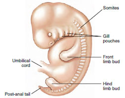 |
| Figure 8-28 Human embryo showing somites, which differentiate into skeletal muscles and axial skeleton. |
Most muscles arise from the mesoderm along each side of the neural tube (Figure 8-28). This mesoderm divides into a linear series of blocklike somites (38 in humans), which by splitting, fusion, and migration become the axial skeleton, dermis of the dorsal skin, and muscles of the back, body wall, and limbs. The limbs begin as buds from the side of the body. Projections of the limb buds develop into fingers and toes.
Mesoderm gives rise to the first functional organ, the embryonic heart. Guided by the underlying endoderm, two clusters of precardiac mesodermal cells move ameba-like into position on either side of the developing gut. These clusters differentiate into a pair of double-walled tubes, which later fuse to form a single, thin tube (see Figure 8-12)
Even while the cells group together, the first twitchings are evident. In a chick embryo, a favorite animal for experimental embryology studies, the primitive heart begins to beat on the second day of the 21-day incubation period; it begins beating before any true blood vessels have formed and before there is any blood to pump. As the ventricle primordium develops, the spon-taneous cellular twitchings become coordinated into a feeble but rhythmical beat. Then, as the atrium develops behind the ventricle, followed by development of the sinus venosus behind the atrium, the heart rate quickens. Each new heart chamber has an intrinsic beat that is faster than its predecessor.
Finally a specialized area of heart muscle called the sinoatrial node develops in the sinus venosus and takes command of the entire heartbeat. The sinoatrial node becomes the heart’s pacemaker. As the heart builds up a strong and efficient beat, vascular channels open within the embryo and across the yolk. Within the vessels are the first primitive blood cells suspended in plasma.
The early development of the heart and circulation is crucial to continued embryonic development, because without a circulation the embryo could not obtain materials for growth. Food is absorbed from the yolk and carried to the embryonic body, oxygen is delivered to all tissues, and carbon dioxide and other wastes are carried away. An embryo is totally dependent on these extraembryonic support systems, and the circulation is the vital link between them.
Support our developers


